3QBK

Bromide-bound form of pharaonis halorhodopsin


Experimental Data Snapshot

  • Method: X-RAY DIFFRACTION
  • Resolution: 2.20 Å
  • R-Value Free: 0.267 
  • R-Value Work: 0.248 
  • R-Value Observed: 0.248 

wwPDB Validation   3D Report Full Report


Ligand Structure Quality Assessment 


This is version 1.2 of the entry. See complete history


Literature

Crystal structures of an O-like blue form and an anion-free yellow form of pharaonis halorhodopsin

Kanada, S.Takeguchi, Y.Murakami, M.Ihara, K.Kouyama, T.

(2011) J Mol Biol 413: 162-176

  • DOI: https://doi.org/10.1016/j.jmb.2011.08.021
  • Primary Citation of Related Structures:  
    3QBG, 3QBI, 3QBK

  • PubMed Abstract: 

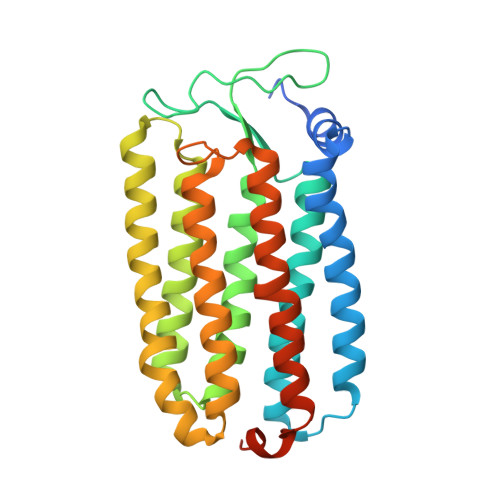
    Halorhodopsin from Natronomonas pharaonis (pHR) was previously crystallized into a monoclinic space group C2, and the structure of the chloride-bound purple form was determined. Here, we report the crystal structures of two chloride-free forms of pHR, that is, an O-like blue form and an M-like yellow form. When the C2 crystal was soaked in a chloride-free alkaline solution, the protein packing was largely altered and the yellow form containing all-trans retinal was generated. Upon neutralization, this yellow form was converted into the blue form. From structural comparison of the different forms of pHR, it was shown that the removal of a chloride ion from the primary binding site (site I), which is located between the retinal Schiff base and Thr126, is accompanied by such a deformation of helix C that the side chain of Thr126 moves toward helix G, leading to a significant shrinkage of site I. A large structural change is also induced in the chloride uptake pathway, where a flip motion of the side chain of Glu234 is accompanied by large movements of the surrounding aromatic residues. Irrespective of different charge distributions at the active site, there was no large difference in the structures of the yellow form and the blue form. It is shown that the yellow-to-purple transition is initiated by the entrance of one water and one HCl to the active site, where the proton and the chloride ion in HCl are transferred to the Schiff base and site I, respectively.


  • Organizational Affiliation

    Department of Physics, Graduate School of Science, Nagoya University, Nagoya 464-8602, Japan.


Macromolecules
Find similar proteins by:  (by identity cutoff)  |  3D Structure
Entity ID: 1
MoleculeChains Sequence LengthOrganismDetailsImage
HalorhodopsinA,
B,
C [auth D]
291Natronomonas pharaonisMutation(s): 0 
Membrane Entity: Yes 
UniProt
Find proteins for P15647 (Natronomonas pharaonis)
Explore P15647 
Go to UniProtKB:  P15647
Entity Groups  
Sequence Clusters30% Identity50% Identity70% Identity90% Identity95% Identity100% Identity
UniProt GroupP15647
Sequence Annotations
Expand
  • Reference Sequence
Small Molecules
Ligands 3 Unique
IDChains Name / Formula / InChI Key2D Diagram3D Interactions
22B
Query on 22B

Download Ideal Coordinates CCD File 
E [auth A],
J [auth B]
BACTERIORUBERIN
C50 H76 O4
UVCQMCCIAHQDAF-CUMPQFAQSA-N
RET
Query on RET

Download Ideal Coordinates CCD File 
D [auth A],
I [auth B],
L [auth D]
RETINAL
C20 H28 O
NCYCYZXNIZJOKI-OVSJKPMPSA-N
BR
Query on BR

Download Ideal Coordinates CCD File 
F [auth A]
G [auth A]
H [auth A]
K [auth B]
M [auth D]
F [auth A],
G [auth A],
H [auth A],
K [auth B],
M [auth D],
N [auth D]
BROMIDE ION
Br
CPELXLSAUQHCOX-UHFFFAOYSA-M
Experimental Data & Validation

Experimental Data

  • Method: X-RAY DIFFRACTION
  • Resolution: 2.20 Å
  • R-Value Free: 0.267 
  • R-Value Work: 0.248 
  • R-Value Observed: 0.248 
  • Space Group: C 1 2 1
Unit Cell:
Length ( Å )Angle ( ˚ )
a = 152.15α = 90
b = 99.72β = 127.8
c = 99.97γ = 90
Software Package:
Software NamePurpose
ADSCdata collection
CNSrefinement
MOSFLMdata reduction
SCALAdata scaling
CNSphasing

Structure Validation

View Full Validation Report


Ligand Structure Quality Assessment 


Entry History 

Deposition Data

Revision History  (Full details and data files)

  • Version 1.0: 2011-08-31
    Type: Initial release
  • Version 1.1: 2013-06-26
    Changes: Database references
  • Version 1.2: 2023-11-01
    Changes: Data collection, Database references, Derived calculations, Refinement description