3QAW

Crystal structure of a glutathione-S-transferase from Antarctic clam Laternula elliptica in a complex with glutathione


Experimental Data Snapshot

  • Method: X-RAY DIFFRACTION
  • Resolution: 2.20 Å
  • R-Value Free: 0.215 
  • R-Value Work: 0.181 

wwPDB Validation   3D Report Full Report


Ligand Structure Quality Assessment 


This is version 1.3 of the entry. See complete history


Literature

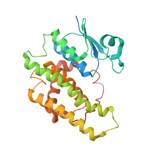
The structure of a shellfish specific GST class glutathione S-transferase from antarctic bivalve Laternula elliptica reveals novel active site architecture.

Park, A.K.Moon, J.H.Jang, E.H.Park, H.Ahn, I.Y.Lee, K.S.Chi, Y.M.

(2013) Proteins 81: 531-537

  • DOI: https://doi.org/10.1002/prot.24208
  • Primary Citation of Related Structures:  
    3QAV, 3QAW

  • PubMed Abstract: 

    Glutathione-S-transferases have been identified in all the living species examined so far, yet little is known about their function in marine organisms. In a previous report, the recently identified GST from Antarctic bivalve Laternula elliptica (LeGST) was classified into the rho class GST, but there are several unique features of LeGST that may justify reclassification, which could represent specific shellfish GSTs. Here, we determined the crystal structure of LeGST, which is a shellfish specific class of GST. The structural analysis showed that the relatively open and wide hydrophobic H-site of the LeGST allows this GST to accommodate various substrates. These results suggest that the H-site of LeGST may be the result of adaptation to their environments as sedentary organisms.


  • Organizational Affiliation

    Division of Biotechnology, College of Life Sciences, Korea University, Seoul 136-713, Republic of Korea.


Macromolecules
Find similar proteins by:  (by identity cutoff)  |  3D Structure
Entity ID: 1
MoleculeChains Sequence LengthOrganismDetailsImage
Rho-class glutathione S-transferase243Laternula ellipticaMutation(s): 0 
EC: 2.5.1.18
UniProt
Find proteins for B9VX79 (Laternula elliptica)
Explore B9VX79 
Go to UniProtKB:  B9VX79
Entity Groups  
Sequence Clusters30% Identity50% Identity70% Identity90% Identity95% Identity100% Identity
UniProt GroupB9VX79
Sequence Annotations
Expand
  • Reference Sequence
Small Molecules
Ligands 1 Unique
IDChains Name / Formula / InChI Key2D Diagram3D Interactions
GSH
Query on GSH

Download Ideal Coordinates CCD File 
B [auth A]GLUTATHIONE
C10 H17 N3 O6 S
RWSXRVCMGQZWBV-WDSKDSINSA-N
Experimental Data & Validation

Experimental Data

  • Method: X-RAY DIFFRACTION
  • Resolution: 2.20 Å
  • R-Value Free: 0.215 
  • R-Value Work: 0.181 
  • Space Group: C 1 2 1
Unit Cell:
Length ( Å )Angle ( ˚ )
a = 90.393α = 90
b = 57.611β = 123.84
c = 55.449γ = 90
Software Package:
Software NamePurpose
CNSrefinement
PDB_EXTRACTdata extraction
HKL-2000data collection
DENZOdata reduction
SCALEPACKdata scaling
HKL-2000data scaling
CNSphasing

Structure Validation

View Full Validation Report


Ligand Structure Quality Assessment 


Entry History 

Deposition Data

Revision History  (Full details and data files)

  • Version 1.0: 2012-02-01
    Type: Initial release
  • Version 1.1: 2013-02-13
    Changes: Database references
  • Version 1.2: 2017-11-08
    Changes: Refinement description
  • Version 1.3: 2023-11-01
    Changes: Data collection, Database references, Derived calculations, Refinement description