3PZP

Human DNA polymerase kappa extending opposite a cis-syn thymine dimer


Experimental Data Snapshot

  • Method: X-RAY DIFFRACTION
  • Resolution: 3.34 Å
  • R-Value Free: 0.288 
  • R-Value Work: 0.242 
  • R-Value Observed: 0.244 

wwPDB Validation   3D Report Full Report


Ligand Structure Quality Assessment 


This is version 1.1 of the entry. See complete history


Literature

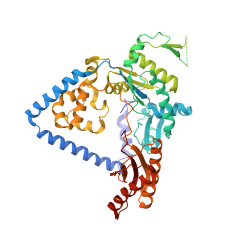
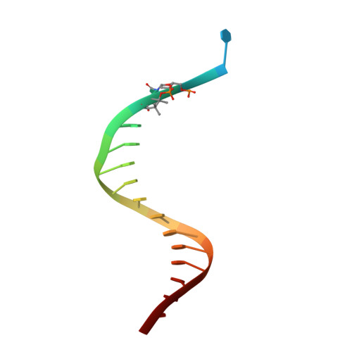
Role of human DNA polymerase kappa in extension opposite from a cis-syn thymine dimer.

Vasquez-Del Carpio, R.Silverstein, T.D.Lone, S.Johnson, R.E.Prakash, L.Prakash, S.Aggarwal, A.K.

(2011) J Mol Biol 408: 252-261

  • DOI: https://doi.org/10.1016/j.jmb.2011.02.042
  • Primary Citation of Related Structures:  
    3PZP

  • PubMed Abstract: 

    Exposure of DNA to UV radiation causes covalent linkages between adjacent pyrimidines. The most common lesion found in DNA from these UV-induced linkages is the cis-syn cyclobutane pyrimidine dimer. Human DNA polymerase κ (Polκ), a member of the Y-family of DNA polymerases, is unable to insert nucleotides opposite the 3'T of a cis-syn T-T dimer, but it can efficiently extend from a nucleotide inserted opposite the 3'T of the dimer by another DNA polymerase. We present here the structure of human Polκ in the act of inserting a nucleotide opposite the 5'T of the cis-syn T-T dimer. The structure reveals a constrained active-site cleft that is unable to accommodate the 3'T of a cis-syn T-T dimer but is remarkably well adapted to accommodate the 5'T via Watson-Crick base pairing, in accord with a proposed role for Polκ in the extension reaction opposite from cyclobutane pyrimidine dimers in vivo.


  • Organizational Affiliation

    Department of Structural and Chemical Biology, Mount Sinai School of Medicine, Box 1677, 1425 Madison Avenue, New York, NY 10029, USA.


Macromolecules

Find similar proteins by:  (by identity cutoff)  |  3D Structure
Entity ID: 1
MoleculeChains Sequence LengthOrganismDetailsImage
DNA polymerase kappa
A, B
517Homo sapiensMutation(s): 0 
Gene Names: POLKDINB1
EC: 2.7.7.7
UniProt & NIH Common Fund Data Resources
Find proteins for Q9UBT6 (Homo sapiens)
Explore Q9UBT6 
Go to UniProtKB:  Q9UBT6
PHAROS:  Q9UBT6
GTEx:  ENSG00000122008 
Entity Groups  
Sequence Clusters30% Identity50% Identity70% Identity90% Identity95% Identity100% Identity
UniProt GroupQ9UBT6
Sequence Annotations
Expand
  • Reference Sequence

Find similar nucleic acids by:  Sequence   |   3D Structure  

Entity ID: 2
MoleculeChains LengthOrganismImage
5'-D(*GP*GP*GP*GP*GP*AP*AP*GP*GP*AP*CP*CP*A)-3'C [auth P],
E [auth Q]
13N/A
Sequence Annotations
Expand
  • Reference Sequence

Find similar nucleic acids by:  Sequence   |   3D Structure  

Entity ID: 3
MoleculeChains LengthOrganismImage
5'-D(*TP*TP*CP*CP*(TTD)P*GP*GP*TP*CP*CP*TP*TP*CP*CP*CP*CP*C)-3'D [auth T],
F [auth U]
17N/A
Sequence Annotations
Expand
  • Reference Sequence
Experimental Data & Validation

Experimental Data

  • Method: X-RAY DIFFRACTION
  • Resolution: 3.34 Å
  • R-Value Free: 0.288 
  • R-Value Work: 0.242 
  • R-Value Observed: 0.244 
  • Space Group: C 2 2 21
Unit Cell:
Length ( Å )Angle ( ˚ )
a = 117.136α = 90
b = 154.476β = 90
c = 217.854γ = 90
Software Package:
Software NamePurpose
REFMACrefinement
PDB_EXTRACTdata extraction
HKL-2000data collection
HKL-2000data reduction
HKL-2000data scaling
PHASERphasing

Structure Validation

View Full Validation Report


Ligand Structure Quality Assessment 


Entry History 

Deposition Data

Revision History  (Full details and data files)

  • Version 1.0: 2011-12-28
    Type: Initial release
  • Version 1.1: 2023-09-13
    Changes: Data collection, Database references, Derived calculations, Refinement description, Structure summary