


Crystal structure of a Nitroreductase with bound FMN (Dhaf_2018) from Desulfitobacterium hafniense DCB-2 at 2.30 A resolution
Joint Center for Structural Genomics (JCSG)To be published.
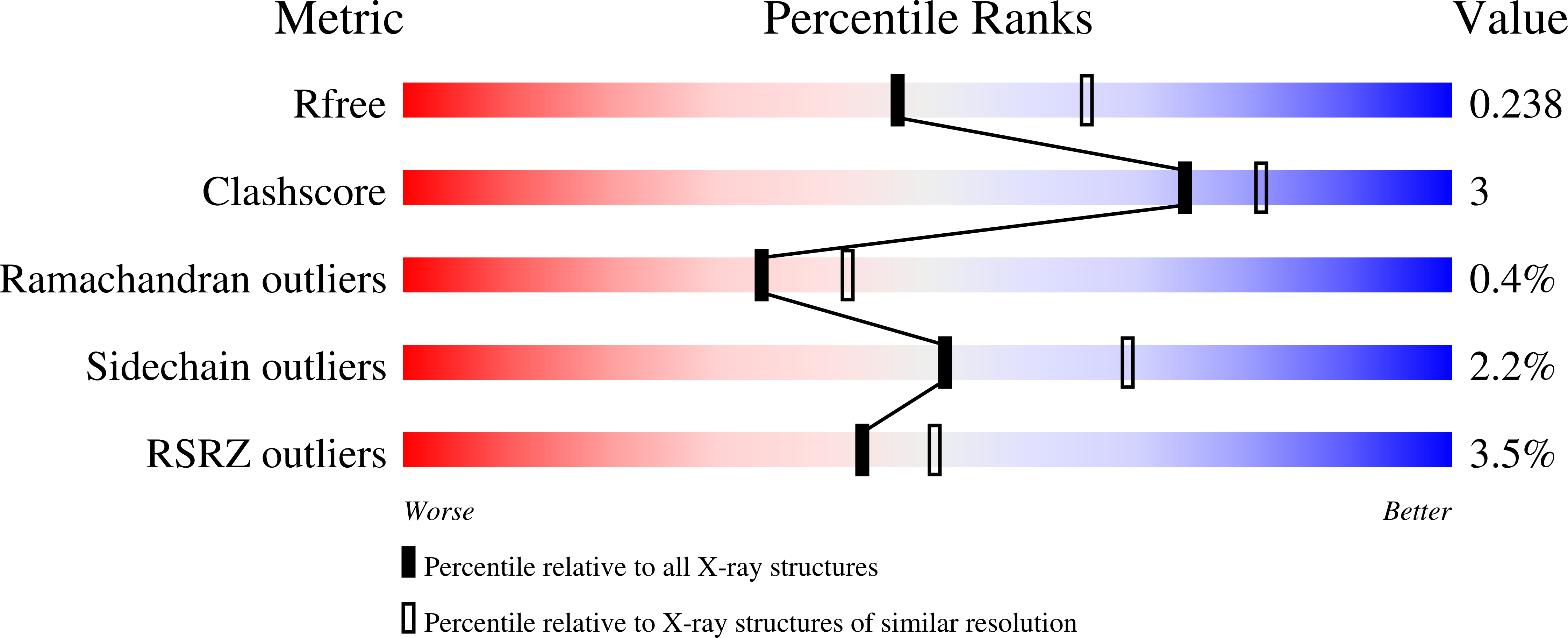
Experimental Data Snapshot
Entity ID: 1 | |||||
|---|---|---|---|---|---|
| Molecule | Chains | Sequence Length | Organism | Details | Image |
| Nitroreductase | 189 | Desulfitobacterium hafniense DCB-2 | Mutation(s): 0 Gene Names: Dhaf_2018, YP_002458491.1 | 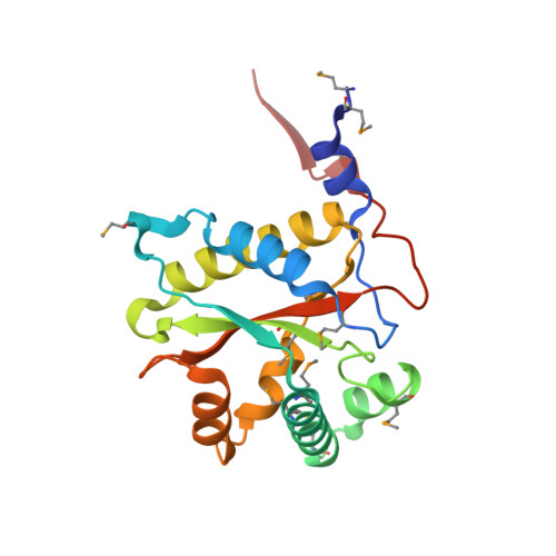 | |
UniProt | |||||
Find proteins for B8FRE0 (Desulfitobacterium hafniense (strain DSM 10664 / DCB-2)) Explore B8FRE0 Go to UniProtKB: B8FRE0 | |||||
Entity Groups | |||||
| Sequence Clusters | 30% Identity50% Identity70% Identity90% Identity95% Identity100% Identity | ||||
| UniProt Group | B8FRE0 | ||||
Sequence AnnotationsExpand | |||||
| |||||
| Ligands 1 Unique | |||||
|---|---|---|---|---|---|
| ID | Chains | Name / Formula / InChI Key | 2D Diagram | 3D Interactions | |
| FMN Query on FMN | E [auth A], F [auth B], G [auth C], H [auth D] | FLAVIN MONONUCLEOTIDE C17 H21 N4 O9 P FVTCRASFADXXNN-SCRDCRAPSA-N |  | ||
| Modified Residues 1 Unique | |||||
|---|---|---|---|---|---|
| ID | Chains | Type | Formula | 2D Diagram | Parent |
| MSE Query on MSE | A, B, C, D | L-PEPTIDE LINKING | C5 H11 N O2 Se |  | MET |
| Length ( Å ) | Angle ( ˚ ) |
|---|---|
| a = 61.261 | α = 90 |
| b = 81.577 | β = 90 |
| c = 161.754 | γ = 90 |
| Software Name | Purpose |
|---|---|
| SHELX | phasing |
| REFMAC | refinement |
| SCALA | data scaling |
| PDB_EXTRACT | data extraction |
| MOSFLM | data reduction |
| SHELXD | phasing |
| autoSHARP | phasing |
RCSB PDB (citation) is hosted by
RCSB PDB is a member of the

