3PPC

Crystal structure of the Candida albicans methionine synthase by surface entropy reduction, tyrosine variant with zinc


Experimental Data Snapshot

  • Method: X-RAY DIFFRACTION
  • Resolution: 2.20 Å
  • R-Value Free: 0.231 
  • R-Value Work: 0.182 
  • R-Value Observed: 0.184 

wwPDB Validation   3D Report Full Report


This is version 1.1 of the entry. See complete history


Literature

Structure of Candida albicans methionine synthase determined by employing surface residue mutagenesis.

Ubhi, D.Kavanagh, K.L.Monzingo, A.F.Robertus, J.D.

(2011) Arch Biochem Biophys 513: 19-26

  • DOI: https://doi.org/10.1016/j.abb.2011.06.002
  • Primary Citation of Related Structures:  
    3PPC, 3PPF, 3PPG, 3PPH

  • PubMed Abstract: 

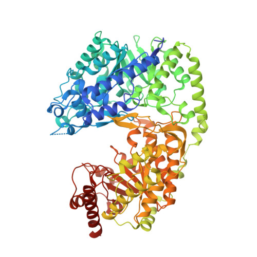
    Fungal methionine synthase, Met6p, transfers a methyl group from 5-methyl-tetrahydrofolate to homocysteine to generate methionine. The enzyme is essential to fungal growth and is a potential anti-fungal drug design target. We have characterized the enzyme from the pathogen Candida albicans but were unable to crystallize it in native form. We converted Lys103, Lys104, and Glu107 all to Tyr (Met6pY), Thr (Met6pT) and Ala (Met6pA). All variants showed wild-type kinetic activity and formed useful crystals, each with unique crystal packing. In each case the mutated residues participated in beneficial crystal contacts. We have solved the three structures at 2.0-2.8Å resolution and analyzed crystal packing, active-site residues, and similarity to other known methionine synthase structures. C. albicans Met6p has a two domain structure with each of the domains having a (βα)(8)-barrel fold. The barrels are arranged face-to-face and the active site is located in a cleft between the two domains. Met6p utilizes a zinc ion for catalysis that is bound in the C-terminal domain and ligated by four conserved residues: His657, Cys659, Glu679 and Cys739.


  • Organizational Affiliation

    Institute for Cellular and Molecular Biology, Department of Chemistry and Biochemistry, University of Texas at Austin, United States.


Macromolecules
Find similar proteins by:  (by identity cutoff)  |  3D Structure
Entity ID: 1
MoleculeChains Sequence LengthOrganismDetailsImage
5-methyltetrahydropteroyltriglutamate--homocysteine methyltransferase
A, B
789Candida albicansMutation(s): 3 
Gene Names: CaO19.10083CaO19.2551MET6
EC: 2.1.1.14
UniProt
Find proteins for P82610 (Candida albicans (strain SC5314 / ATCC MYA-2876))
Explore P82610 
Go to UniProtKB:  P82610
Entity Groups  
Sequence Clusters30% Identity50% Identity70% Identity90% Identity95% Identity100% Identity
UniProt GroupP82610
Sequence Annotations
Expand
  • Reference Sequence
Small Molecules
Ligands 2 Unique
IDChains Name / Formula / InChI Key2D Diagram3D Interactions
ZN
Query on ZN

Download Ideal Coordinates CCD File 
C [auth A],
I [auth B]
ZINC ION
Zn
PTFCDOFLOPIGGS-UHFFFAOYSA-N
CL
Query on CL

Download Ideal Coordinates CCD File 
D [auth A]
E [auth A]
F [auth A]
G [auth A]
H [auth A]
D [auth A],
E [auth A],
F [auth A],
G [auth A],
H [auth A],
J [auth B],
K [auth B],
L [auth B]
CHLORIDE ION
Cl
VEXZGXHMUGYJMC-UHFFFAOYSA-M
Experimental Data & Validation

Experimental Data

  • Method: X-RAY DIFFRACTION
  • Resolution: 2.20 Å
  • R-Value Free: 0.231 
  • R-Value Work: 0.182 
  • R-Value Observed: 0.184 
  • Space Group: P 21 21 21
Unit Cell:
Length ( Å )Angle ( ˚ )
a = 77.676α = 90
b = 92.685β = 90
c = 190.97γ = 90
Software Package:
Software NamePurpose
SCALAdata scaling
REFMACrefinement
PDB_EXTRACTdata extraction
ADSCdata collection
MOSFLMdata reduction
PHASERphasing

Structure Validation

View Full Validation Report


Entry History 

Deposition Data

Revision History  (Full details and data files)

  • Version 1.0: 2011-10-12
    Type: Initial release
  • Version 1.1: 2023-09-06
    Changes: Data collection, Database references, Derived calculations, Refinement description