3POR

PORIN CONFORMATION IN THE ABSENCE OF CALCIUM; REFINED STRUCTURE AT 2.5 ANGSTROMS RESOLUTION


Experimental Data Snapshot

  • Method: X-RAY DIFFRACTION
  • Resolution: 2.50 Å
  • R-Value Work: 0.188 

wwPDB Validation   3D Report Full Report


Ligand Structure Quality Assessment 


This is version 1.5 of the entry. See complete history


Literature

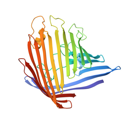
Porin conformation in the absence of calcium. Refined structure at 2.5 A resolution.

Weiss, M.S.Schulz, G.E.

(1993) J Mol Biol 231: 817-824

  • DOI: https://doi.org/10.1006/jmbi.1993.1328
  • Primary Citation of Related Structures:  
    3POR

  • PubMed Abstract: 

    The crystal structure of porin from Rhodobacter capsulatus in the absence of divalent calcium ions has been refined to convergence at a resolution of 2.5 A using the simulated annealing refinement method. The final model consists of all 301 amino acid residues, 77 solvent molecules, one tris(hydroxymethyl)-aminomethane molecule and one unknown ligand modeled as n-octyltetraoxyethylene. A superposition with the previously described model containing three calcium ions showed structural changes at the segment 108-116 of the inner loop beta 5-beta 6, and at loops beta 8-beta 9 and beta 11-beta 12 at the extracellular side of the porin molecule. Evidence is presented that the conformational changes depend on the presence or absence of calcium ions. A possible influence on porin function is discussed.


  • Organizational Affiliation

    Institut für Organische Chemie und Biochemie, Albert-Ludwigs-Universität, Freiburg im Breisgau, Germany.


Macromolecules
Find similar proteins by:  (by identity cutoff)  |  3D Structure
Entity ID: 1
MoleculeChains Sequence LengthOrganismDetailsImage
PORIN301Rhodobacter capsulatusMutation(s): 0 
Membrane Entity: Yes 
UniProt
Find proteins for P31243 (Rhodobacter capsulatus)
Explore P31243 
Go to UniProtKB:  P31243
Entity Groups  
Sequence Clusters30% Identity50% Identity70% Identity90% Identity95% Identity100% Identity
UniProt GroupP31243
Sequence Annotations
Expand
  • Reference Sequence
Small Molecules
Ligands 2 Unique
IDChains Name / Formula / InChI Key2D Diagram3D Interactions
C8E
Query on C8E

Download Ideal Coordinates CCD File 
B [auth A](HYDROXYETHYLOXY)TRI(ETHYLOXY)OCTANE
C16 H34 O5
FEOZZFHAVXYAMB-UHFFFAOYSA-N
TRS
Query on TRS

Download Ideal Coordinates CCD File 
C [auth A]2-AMINO-2-HYDROXYMETHYL-PROPANE-1,3-DIOL
C4 H12 N O3
LENZDBCJOHFCAS-UHFFFAOYSA-O
Experimental Data & Validation

Experimental Data

  • Method: X-RAY DIFFRACTION
  • Resolution: 2.50 Å
  • R-Value Work: 0.188 
  • Space Group: H 3
Unit Cell:
Length ( Å )Angle ( ˚ )
a = 95.3α = 90
b = 95.3β = 90
c = 146.8γ = 120
Software Package:
Software NamePurpose
X-PLORrefinement

Structure Validation

View Full Validation Report


Ligand Structure Quality Assessment 


Entry History 

Deposition Data

Revision History  (Full details and data files)

  • Version 1.0: 1993-07-15
    Type: Initial release
  • Version 1.1: 2008-03-25
    Changes: Version format compliance
  • Version 1.2: 2011-07-13
    Changes: Derived calculations, Version format compliance
  • Version 1.3: 2011-11-16
    Changes: Atomic model
  • Version 1.4: 2017-11-29
    Changes: Derived calculations, Other
  • Version 1.5: 2024-02-21
    Changes: Data collection, Database references, Derived calculations