


3PL3
Crystal structure of Cel7A from Talaromyces emersonii in complex with cellopentaose
- PDB DOI: https://doi.org/10.2210/pdb3PL3/pdb
- Classification: HYDROLASE
- Organism(s): Rasamsonia emersonii
- Expression System: Aspergillus niger
- Mutation(s): No
- Deposited: 2010-11-12 Released: 2011-12-14
Experimental Data Snapshot
- Method: X-RAY DIFFRACTION
- Resolution: 1.18 Å
- R-Value Free: 0.150
- R-Value Work: 0.127
- R-Value Observed: 0.127
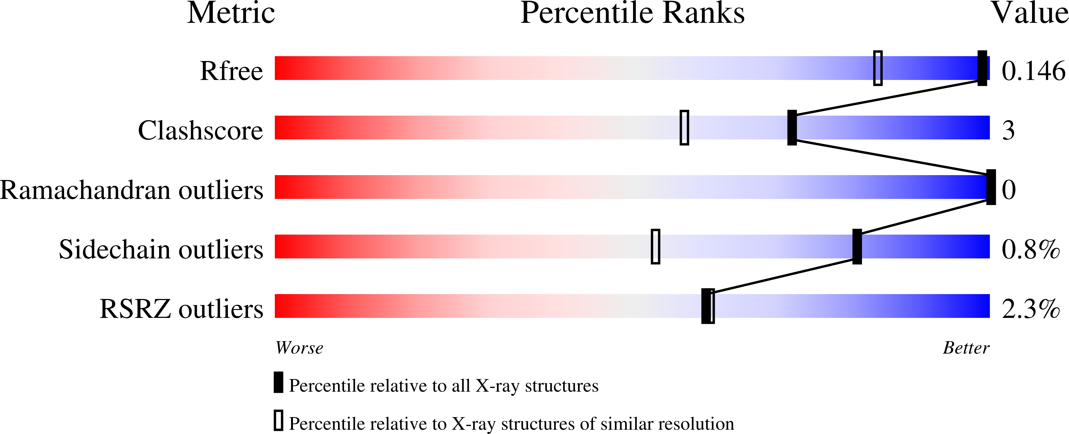
wwPDB Validation 3D Report Full Report
This is version 4.1 of the entry. See complete history.

Macromolecules
Find similar proteins by:
(by identity cutoff) | 3D Structure
Entity ID: 1 | |||||
|---|---|---|---|---|---|
| Molecule | Chains | Sequence Length | Organism | Details | Image |
| Cellobiohydrolase 1 catalytic domain | 437 | Rasamsonia emersonii | Mutation(s): 0 Gene Names: cbh1, cbh1A EC: 3.2.1.91 | 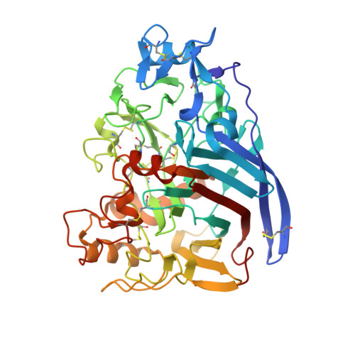 | |
UniProt | |||||
Find proteins for Q8TFL9 (Talaromyces emersonii) Explore Q8TFL9 Go to UniProtKB: Q8TFL9 | |||||
Entity Groups | |||||
| Sequence Clusters | 30% Identity50% Identity70% Identity90% Identity95% Identity100% Identity | ||||
| UniProt Group | Q8TFL9 | ||||
Sequence AnnotationsExpand | |||||
| |||||
Oligosaccharides
Entity ID: 2 | |||||
|---|---|---|---|---|---|
| Molecule | Chains | Length | 2D Diagram | Glycosylation | 3D Interactions |
| beta-D-mannopyranose-(1-4)-2-acetamido-2-deoxy-beta-D-glucopyranose-(1-4)-2-acetamido-2-deoxy-beta-D-glucopyranose | B | 3 |  | N-Glycosylation | |
Glycosylation Resources | |||||
GlyTouCan: G15407YE GlyCosmos: G15407YE GlyGen: G15407YE | |||||
Entity ID: 3 | |||||
|---|---|---|---|---|---|
| Molecule | Chains | Length | 2D Diagram | Glycosylation | 3D Interactions |
| 2-acetamido-2-deoxy-beta-D-glucopyranose-(1-4)-2-acetamido-2-deoxy-beta-D-glucopyranose | C | 2 |  | N-Glycosylation | |
Glycosylation Resources | |||||
GlyTouCan: G42666HT GlyCosmos: G42666HT GlyGen: G42666HT | |||||


Small Molecules
| Ligands 1 Unique | |||||
|---|---|---|---|---|---|
| ID | Chains | Name / Formula / InChI Key | 2D Diagram | 3D Interactions | |
| SO4 Query on SO4 | F [auth A] | SULFATE ION O4 S QAOWNCQODCNURD-UHFFFAOYSA-L |  | ||
| Modified Residues 1 Unique | |||||
|---|---|---|---|---|---|
| ID | Chains | Type | Formula | 2D Diagram | Parent |
| PCA Query on PCA | A | L-PEPTIDE LINKING | C5 H7 N O3 |  | GLN |
Biologically Interesting Molecules (External Reference) 2 Unique
Entity ID: 4 | |||||
|---|---|---|---|---|---|
| ID | Chains | Name | Type/Class | 2D Diagram | 3D Interactions |
| PRD_900016 Query on PRD_900016 | D | beta-cellopentaose | Oligosaccharide / Metabolism |  | |
Entity ID: 5 | |||||
|---|---|---|---|---|---|
| ID | Chains | Name | Type/Class | 2D Diagram | 3D Interactions |
| PRD_900021 Query on PRD_900021 | E | beta-cellotriose | Oligosaccharide / Metabolism |  | |
Experimental Data & Validation
Experimental Data
- Method: X-RAY DIFFRACTION
- Resolution: 1.18 Å
- R-Value Free: 0.150
- R-Value Work: 0.127
- R-Value Observed: 0.127
- Space Group: P 41 21 2
Unit Cell:
| Length ( Å ) | Angle ( ˚ ) |
|---|---|
| a = 74.204 | α = 90 |
| b = 74.204 | β = 90 |
| c = 175.457 | γ = 90 |
| Software Name | Purpose |
|---|---|
| BOS | data collection |
| PHASER | phasing |
| PHENIX | refinement |
| HKL-2000 | data reduction |
| HKL-2000 | data scaling |
Entry History
Deposition Data
- Released Date: 2011-12-14 Deposition Author(s): Qin, L., Pereira, J.H., McAndrew, R.P., Simmons, B.A., Sapra, R., Adams, P.D., Sale, K.L.
Revision History (Full details and data files)
- Version 1.0: 2011-12-14
Type: Initial release - Version 2.0: 2019-12-25
Changes: Derived calculations, Polymer sequence - Version 3.0: 2020-07-29
Type: Remediation
Reason: Carbohydrate remediation
Changes: Advisory, Atomic model, Data collection, Derived calculations, Non-polymer description, Structure summary - Version 4.0: 2020-10-14
Changes: Atomic model, Structure summary - Version 4.1: 2023-09-06
Changes: Data collection, Database references, Refinement description
