


Structure and stability of M.jannaschii L7Ae El9 KtoQ mutant
Biswas, S., Gagnon, K.T., Mattos, C., Brown, B.A., Maxwell, E.S.To be published.
Experimental Data Snapshot
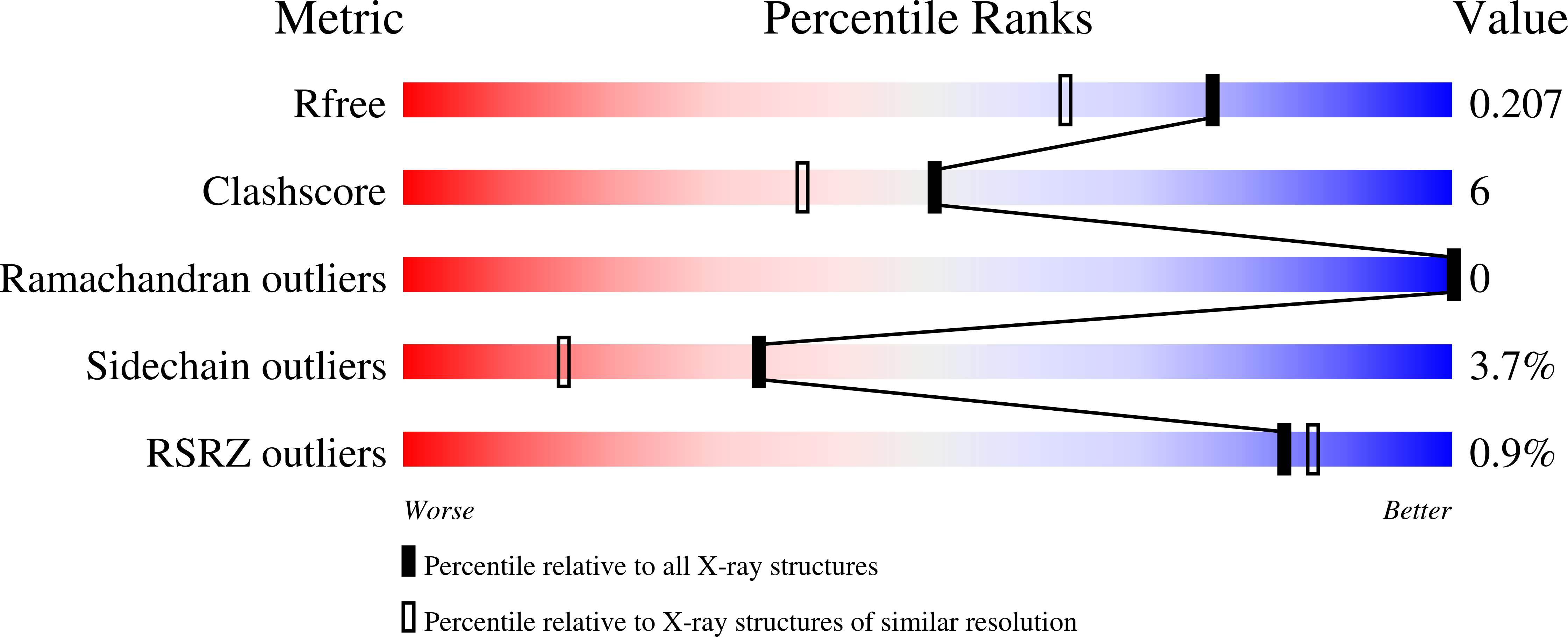
wwPDB Validation 3D Report Full Report
Entity ID: 1 | |||||
|---|---|---|---|---|---|
| Molecule | Chains | Sequence Length | Organism | Details | Image |
| 50S ribosomal protein L7Ae | 117 | Methanocaldococcus jannaschii | Mutation(s): 5 Gene Names: rpl7ae, MJ1203 | 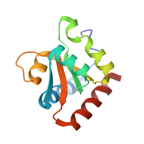 | |
UniProt | |||||
Find proteins for P54066 (Methanocaldococcus jannaschii (strain ATCC 43067 / DSM 2661 / JAL-1 / JCM 10045 / NBRC 100440)) Explore P54066 Go to UniProtKB: P54066 | |||||
Entity Groups | |||||
| Sequence Clusters | 30% Identity50% Identity70% Identity90% Identity95% Identity100% Identity | ||||
| UniProt Group | P54066 | ||||
Sequence AnnotationsExpand | |||||
| |||||
| Ligands 2 Unique | |||||
|---|---|---|---|---|---|
| ID | Chains | Name / Formula / InChI Key | 2D Diagram | 3D Interactions | |
| SO4 Query on SO4 | C [auth A], D [auth A] | SULFATE ION O4 S QAOWNCQODCNURD-UHFFFAOYSA-L |  | ||
| ACT Query on ACT | E [auth B], F [auth B] | ACETATE ION C2 H3 O2 QTBSBXVTEAMEQO-UHFFFAOYSA-M |  | ||
| Length ( Å ) | Angle ( ˚ ) |
|---|---|
| a = 43.443 | α = 90 |
| b = 55.634 | β = 90 |
| c = 103.039 | γ = 90 |
| Software Name | Purpose |
|---|---|
| SERGUI | data collection |
| PHASER | phasing |
| PHENIX | refinement |
| HKL-2000 | data reduction |
| HKL-2000 | data scaling |
RCSB PDB (citation) is hosted by
RCSB PDB is a member of the

