3P6B

The crystal structure of CelK CBM4 from Clostridium thermocellum


Experimental Data Snapshot

  • Method: X-RAY DIFFRACTION
  • Resolution: 2.00 Å
  • R-Value Free: 0.274 
  • R-Value Work: 0.212 
  • R-Value Observed: 0.215 

wwPDB Validation   3D Report Full Report


This is version 1.2 of the entry. See complete history


Literature

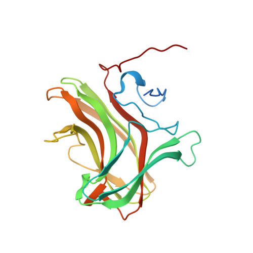
Structure of CBM4 from Clostridium thermocellum cellulase K.

Alahuhta, M.Luo, Y.Ding, S.Y.Himmel, M.E.Lunin, V.V.

(2011) Acta Crystallogr Sect F Struct Biol Cryst Commun 67: 527-530

  • DOI: https://doi.org/10.1107/S1744309111003307
  • Primary Citation of Related Structures:  
    3P6B

  • PubMed Abstract: 

    Here, a 2.0 Å resolution X-ray structure of Clostridium thermocellum cellulase K family 4 carbohydrate-binding module (CelK CBM4) is reported. The resulting structure was refined to an R factor of 0.212 and an R(free) of 0.274. Structural analysis shows that this new structure is very similar to the previously solved structure of C. thermocellum CbhA CBM4. Most importantly, these data support the previously proposed notion of an extended binding pocket using a novel tryptophan-containing loop that may be highly conserved in clostridial CBM4 proteins.


  • Organizational Affiliation

    BioSciences Center, National Renewable Energy Laboratory, 1617 Cole Boulevard, Golden, Colorado 80401-3305, USA.


Macromolecules
Find similar proteins by:  (by identity cutoff)  |  3D Structure
Entity ID: 1
MoleculeChains Sequence LengthOrganismDetailsImage
Cellulose 1,4-beta-cellobiosidase
A, B
205Acetivibrio thermocellusMutation(s): 0 
Gene Names: celK
EC: 3.2.1.91
UniProt
Find proteins for P0C2S1 (Acetivibrio thermocellus)
Explore P0C2S1 
Go to UniProtKB:  P0C2S1
Entity Groups  
Sequence Clusters30% Identity50% Identity70% Identity90% Identity95% Identity100% Identity
UniProt GroupP0C2S1
Sequence Annotations
Expand
  • Reference Sequence
Small Molecules
Ligands 4 Unique
IDChains Name / Formula / InChI Key2D Diagram3D Interactions
SO4
Query on SO4

Download Ideal Coordinates CCD File 
H [auth A]
I [auth A]
J [auth A]
K [auth A]
N [auth B]
H [auth A],
I [auth A],
J [auth A],
K [auth A],
N [auth B],
O [auth B],
P [auth B],
Q [auth B],
R [auth B]
SULFATE ION
O4 S
QAOWNCQODCNURD-UHFFFAOYSA-L
GOL
Query on GOL

Download Ideal Coordinates CCD File 
G [auth A],
M [auth B]
GLYCEROL
C3 H8 O3
PEDCQBHIVMGVHV-UHFFFAOYSA-N
ACT
Query on ACT

Download Ideal Coordinates CCD File 
D [auth A],
E [auth A],
F [auth A]
ACETATE ION
C2 H3 O2
QTBSBXVTEAMEQO-UHFFFAOYSA-M
CA
Query on CA

Download Ideal Coordinates CCD File 
C [auth A],
L [auth B]
CALCIUM ION
Ca
BHPQYMZQTOCNFJ-UHFFFAOYSA-N
Experimental Data & Validation

Experimental Data

  • Method: X-RAY DIFFRACTION
  • Resolution: 2.00 Å
  • R-Value Free: 0.274 
  • R-Value Work: 0.212 
  • R-Value Observed: 0.215 
  • Space Group: P 41 21 2
Unit Cell:
Length ( Å )Angle ( ˚ )
a = 65.949α = 90
b = 65.949β = 90
c = 272.436γ = 90
Software Package:
Software NamePurpose
PROTEUM PLUSdata collection
MOLREPphasing
REFMACrefinement
SAINTdata reduction
PROTEUM PLUSdata scaling

Structure Validation

View Full Validation Report


Entry History 

Deposition Data

Revision History  (Full details and data files)

  • Version 1.0: 2011-08-24
    Type: Initial release
  • Version 1.1: 2013-03-27
    Changes: Other
  • Version 1.2: 2023-09-06
    Changes: Data collection, Database references, Derived calculations, Refinement description