Structural evidence for stabilization of inhibitor binding by a protein cavity in the dehaloperoxidase-hemoglobin from Amphitrite ornata.
de Serrano, V., Franzen, S.(2012) Biopolymers 98: 27-35
- PubMed: 23325557
- DOI: https://doi.org/10.1002/bip.21674
- Primary Citation of Related Structures:
3ORD - PubMed Abstract:
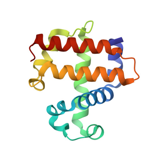
A functional role for a protein cavity that stabilizes inhibitor binding has been established based on a comparison of Xe-derivatized and inhibitor-bound X-ray crystal structures in dehaloperoxidase-hemoglobin (DHP A) of Amphitrite ornata. The internal binding affinity of four different inhibitors, 4-fluorophenol, 4-chlorophenol, 4-bromophenol, and 4-iodophenol in the distal pocket has been shown previously to increase proportional to the radius of the para-halogen atom. Inhibition of oxidation of the native substrate, 2,4,6-tribromophenol, has been shown to follow the trend in inhibitor binding strength, because of a two-site competitive inhibition mechanism that involves displacement of the substrate by the inhibitor in a gated mechanism involving the distal histidine of DHP A. In this study, it is shown that the origin of the stronger binding by a larger para-halogen substituent coincides structurally with a Xe-binding cavity (Xe1) characterized structurally by X-ray crystallography. The Xe1 site is surrounded by amino acid resides L100, F21, F24, F35, F60, and V59 in the distal pocket, located 4.8 Å from the heme iron, in a position that is coincident with the para-bromine atom of the inhibitor 4-bromophenol. 4-bromophenol is prevalent in benthic ecosystems where A. ornata resides. A second, less well-defined, binding site in DHP A, labeled as Xe2, is located near the surface of the protein in the vicinity of amino acid residues L62, R69, D79, T82, and L83, which may be related to substrate docking on the surface of DHP A.
Organizational Affiliation:
Department of Chemistry, North Carolina State University, Raleigh, NC 27695.