3OD9

Crystal structure of PliI-Ah, periplasmic lysozyme inhibitor of I-type lysozyme from Aeromonas hydrophyla


Experimental Data Snapshot

  • Method: X-RAY DIFFRACTION
  • Resolution: 1.41 Å
  • R-Value Free: 0.197 
  • R-Value Work: 0.161 
  • R-Value Observed: 0.163 

wwPDB Validation   3D Report Full Report


This is version 1.2 of the entry. See complete history


Literature

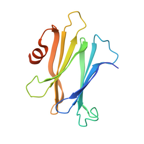
Molecular Basis of Bacterial Defense against Host Lysozymes: X-ray Structures of Periplasmic Lysozyme Inhibitors PliI and PliC.

Leysen, S.Van Herreweghe, J.M.Callewaert, L.Heirbaut, M.Buntinx, P.Michiels, C.W.Strelkov, S.V.

(2011) J Mol Biol 405: 1233-1245

  • DOI: https://doi.org/10.1016/j.jmb.2010.12.007
  • Primary Citation of Related Structures:  
    3OD9, 3OE3

  • PubMed Abstract: 

    Lysozymes play a key role in the innate immune system of vertebrates and invertebrates by hydrolyzing peptidoglycan, a vital component of the bacterial cell wall. Gram-negative bacteria produce various types of lysozyme inhibitors that allow them to survive the bactericidal action of lysozyme when their outer membrane is permeabilized. So far, three lysozyme inhibitor families have been described: the Ivy (inhibitor of vertebrate lysozyme) family, the MliC/PliC (membrane-associated/periplasmic lysozyme inhibitor of C-type lysozyme) family, and the PliI (periplasmic lysozyme inhibitor of I-type lysozyme) family. Here, we report high-resolution crystal structures of Salmonella typhimurium PliC (PliC-St) and Aeromonas hydrophila PliI (PliI-Ah). The structure of PliI-Ah is the first in the recently discovered PliI family of lysozyme inhibitors, while the structure of PliC-St is the first structure of a periplasmic lysozyme inhibitor from the PliC/MliC family. Using small-angle X-ray scattering, we demonstrate that both PliC-St and PliI-Ah form stable dimers in solution. The functional dimer architecture of PliC-St is very different from that of the recently described MliC from Pseudomonas aeruginosa (MliC-Pa), despite the close resemblance of their monomers. Furthermore, PliI-Ah has distinctly different monomer and dimer folds compared to PliC, MliC, and Ivy proteins. Site-directed mutagenesis suggests that the inhibitory action of PliI-Ah proceeds via an insertion of a loop containing the conserved SGxY motif into the active center of I-type lysozymes. This motif is related to the functional SGxxY motif found in the MliC/PliC family.


  • Organizational Affiliation

    Laboratory for Biocrystallography, Department of Pharmaceutical Sciences, Katholieke Universiteit Leuven, Herestraat 49 bus 822, 3000 Leuven, Belgium.


Macromolecules
Find similar proteins by:  (by identity cutoff)  |  3D Structure
Entity ID: 1
MoleculeChains Sequence LengthOrganismDetailsImage
Putative exported protein
A, B, C
135Aeromonas hydrophila subsp. hydrophila ATCC 7966Mutation(s): 0 
Gene Names: AHA_1205
UniProt
Find proteins for A0KHJ5 (Aeromonas hydrophila subsp. hydrophila (strain ATCC 7966 / DSM 30187 / BCRC 13018 / CCUG 14551 / JCM 1027 / KCTC 2358 / NCIMB 9240 / NCTC 8049))
Explore A0KHJ5 
Go to UniProtKB:  A0KHJ5
Entity Groups  
Sequence Clusters30% Identity50% Identity70% Identity90% Identity95% Identity100% Identity
UniProt GroupA0KHJ5
Sequence Annotations
Expand
  • Reference Sequence
Small Molecules
Ligands 2 Unique
IDChains Name / Formula / InChI Key2D Diagram3D Interactions
K
Query on K

Download Ideal Coordinates CCD File 
D [auth A],
K [auth B]
POTASSIUM ION
K
NPYPAHLBTDXSSS-UHFFFAOYSA-N
NA
Query on NA

Download Ideal Coordinates CCD File 
E [auth A]
F [auth A]
G [auth A]
H [auth A]
I [auth A]
E [auth A],
F [auth A],
G [auth A],
H [auth A],
I [auth A],
J [auth A],
L [auth B],
M [auth C],
N [auth C],
O [auth C]
SODIUM ION
Na
FKNQFGJONOIPTF-UHFFFAOYSA-N
Experimental Data & Validation

Experimental Data

  • Method: X-RAY DIFFRACTION
  • Resolution: 1.41 Å
  • R-Value Free: 0.197 
  • R-Value Work: 0.161 
  • R-Value Observed: 0.163 
  • Space Group: C 1 2 1
Unit Cell:
Length ( Å )Angle ( ˚ )
a = 103.021α = 90
b = 60.327β = 115.65
c = 82.187γ = 90
Software Package:
Software NamePurpose
SCALAdata scaling
PHENIXrefinement
PDB_EXTRACTdata extraction
XSCALEdata scaling
MOLREPphasing

Structure Validation

View Full Validation Report


Entry History 

Deposition Data

Revision History  (Full details and data files)

  • Version 1.0: 2010-12-22
    Type: Initial release
  • Version 1.1: 2011-07-13
    Changes: Version format compliance
  • Version 1.2: 2024-02-21
    Changes: Data collection, Database references, Derived calculations