3OBA

Structure of the beta-galactosidase from Kluyveromyces lactis


Experimental Data Snapshot

  • Method: X-RAY DIFFRACTION
  • Resolution: 2.75 Å
  • R-Value Free: 0.243 
  • R-Value Work: 0.207 
  • R-Value Observed: 0.208 

wwPDB Validation   3D Report Full Report


This is version 1.3 of the entry. See complete history


Literature

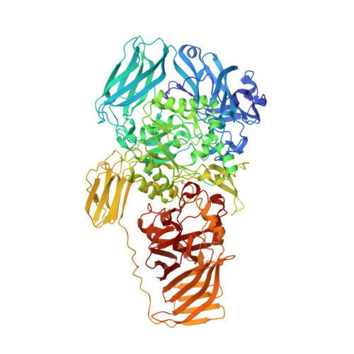
Structural basis of specificity in tetrameric Kluyveromyces lactis beta-galactosidase.

Pereira-Rodriguez, A.Fernandez-Leiro, R.Gonzalez-Siso, M.I.Cerdan, M.E.Becerra, M.Sanz-Aparicio, J.

(2012) J Struct Biol 177: 392-401

  • DOI: https://doi.org/10.1016/j.jsb.2011.11.031
  • Primary Citation of Related Structures:  
    3OB8, 3OBA

  • PubMed Abstract: 

    β-Galactosidase or lactase is a very important enzyme in the food industry, being that from the yeast Kluyveromyces lactis the most widely used. Here we report its three-dimensional structure both in the free state and complexed with the product galactose. The monomer folds into five domains in a pattern conserved with the prokaryote enzymes of the GH2 family, although two long insertions in domains 2 and 3 are unique and related to oligomerization and specificity. The tetrameric enzyme is a dimer of dimers, with higher dissociation energy for the dimers than for its assembly. Two active centers are located at the interface within each dimer in a narrow channel. The insertion at domain 3 protrudes into this channel and makes putative links with the aglycone moiety of docked lactose. In spite of common structural features related to function, the determinants of the reaction mechanism proposed for Escherichia coli β-galactosidase are not found in the active site of the K. lactis enzyme. This is the first X-ray crystal structure for a β-galactosidase used in food processing.


  • Organizational Affiliation

    Departamento de Bioloxía Celular e Molecular, Facultade de Ciencias, Universidade da Coruña, Campus da Zapateira s/n, 15071-A Coruña, Spain. apereira@udc.es


Macromolecules
Find similar proteins by:  (by identity cutoff)  |  3D Structure
Entity ID: 1
MoleculeChains Sequence LengthOrganismDetailsImage
Beta-galactosidaseA,
B [auth C],
C [auth D],
D [auth B]
1,032Kluyveromyces lactisMutation(s): 0 
Gene Names: KLLA0B14883gLAC4
EC: 3.2.1.23
UniProt
Find proteins for P00723 (Kluyveromyces lactis (strain ATCC 8585 / CBS 2359 / DSM 70799 / NBRC 1267 / NRRL Y-1140 / WM37))
Explore P00723 
Go to UniProtKB:  P00723
Entity Groups  
Sequence Clusters30% Identity50% Identity70% Identity90% Identity95% Identity100% Identity
UniProt GroupP00723
Sequence Annotations
Expand
  • Reference Sequence
Small Molecules
Ligands 2 Unique
IDChains Name / Formula / InChI Key2D Diagram3D Interactions
GOL
Query on GOL

Download Ideal Coordinates CCD File 
F [auth A]
G [auth A]
I [auth C]
J [auth C]
K [auth C]
F [auth A],
G [auth A],
I [auth C],
J [auth C],
K [auth C],
L [auth C],
N [auth D],
O [auth D],
Q [auth B],
R [auth B]
GLYCEROL
C3 H8 O3
PEDCQBHIVMGVHV-UHFFFAOYSA-N
MN3
Query on MN3

Download Ideal Coordinates CCD File 
E [auth A],
H [auth C],
M [auth D],
P [auth B]
MANGANESE (III) ION
Mn
MMIPFLVOWGHZQD-UHFFFAOYSA-N
Experimental Data & Validation

Experimental Data

  • Method: X-RAY DIFFRACTION
  • Resolution: 2.75 Å
  • R-Value Free: 0.243 
  • R-Value Work: 0.207 
  • R-Value Observed: 0.208 
  • Space Group: P 21 21 21
Unit Cell:
Length ( Å )Angle ( ˚ )
a = 140.03α = 90
b = 153.34β = 90
c = 216.16γ = 90
Software Package:
Software NamePurpose
MOSFLMdata reduction
SCALAdata scaling
MOLREPphasing
REFMACrefinement
PDB_EXTRACTdata extraction
ADSCdata collection

Structure Validation

View Full Validation Report


Entry History 

Deposition Data

Revision History  (Full details and data files)

  • Version 1.0: 2011-08-17
    Type: Initial release
  • Version 1.1: 2012-02-08
    Changes: Database references
  • Version 1.2: 2013-06-26
    Changes: Database references
  • Version 1.3: 2023-09-06
    Changes: Data collection, Database references, Derived calculations, Refinement description