3NF9

Structural basis for a new mechanism of inhibition of HIV integrase identified by fragment screening and structure based design


Experimental Data Snapshot

  • Method: X-RAY DIFFRACTION
  • Resolution: 1.95 Å
  • R-Value Free: 0.208 
  • R-Value Work: 0.164 
  • R-Value Observed: 0.166 

wwPDB Validation   3D Report Full Report


Ligand Structure Quality Assessment 


This is version 1.3 of the entry. See complete history


Literature

Structural basis for a new mechanism of inhibition of HIV-1 integrase identified by fragment screening and structure-based design

Rhodes, D.I.Peat, T.S.Vandegraaff, N.Jeevarajah, D.Le, G.Jones, E.D.Smith, J.A.Coates, J.A.Winfield, L.J.Thienthong, N.Newman, J.Lucent, D.Ryan, J.H.Savage, G.P.Francis, C.L.Deadman, J.J.

(2011) Antivir Chem Chemother 21: 155-168

  • DOI: https://doi.org/10.3851/IMP1716
  • Primary Citation of Related Structures:  
    3NF6, 3NF7, 3NF8, 3NF9, 3NFA

  • PubMed Abstract: 

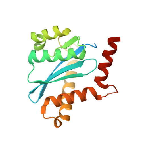
    HIV-1 integrase is a clinically validated therapeutic target for the treatment of HIV-1 infection, with one approved therapeutic currently on the market. This enzyme represents an attractive target for the development of new inhibitors to HIV-1 that are effective against the current resistance mutations. A fragment-based screening method employing surface plasmon resonance and NMR was initially used to detect interactions between integrase and fragments. The binding sites of the fragments were elucidated by crystallography and the structural information used to design and synthesize improved ligands. The location of binding of fragments to the catalytic core of integrase was found to be in a previously undescribed binding site, adjacent to the mobile loop. Enzyme assays confirmed that formation of enzyme-fragment complexes inhibits the catalytic activity of integrase and the structural data was utilized to further develop these fragments into more potent novel enzyme inhibitors. We have defined a new site in integrase as a valid region for the structure-based design of allosteric integrase inhibitors. Using a structure-based design process we have improved the activity of the initial fragments 45-fold.


  • Organizational Affiliation

    Avexa Ltd, Richmond, Australia. drhodes@jdjbioservices.com


Macromolecules
Find similar proteins by:  (by identity cutoff)  |  3D Structure
Entity ID: 1
MoleculeChains Sequence LengthOrganismDetailsImage
Integrase
A, B
183Human immunodeficiency virus 1Mutation(s): 3 
UniProt
Find proteins for P12497 (Human immunodeficiency virus type 1 group M subtype B (isolate NY5))
Explore P12497 
Go to UniProtKB:  P12497
Entity Groups  
Sequence Clusters30% Identity50% Identity70% Identity90% Identity95% Identity100% Identity
UniProt GroupP12497
Sequence Annotations
Expand
  • Reference Sequence
Small Molecules
Ligands 4 Unique
IDChains Name / Formula / InChI Key2D Diagram3D Interactions
CD9
Query on CD9

Download Ideal Coordinates CCD File 
H [auth A],
S [auth B]
5-[(6-chloro-2-oxo-2,3-dihydro-1H-indol-1-yl)methyl]-1,3-benzodioxole-4-carboxylic acid
C17 H12 Cl N O5
XVKFXTJLVSDWCE-UHFFFAOYSA-N
SO4
Query on SO4

Download Ideal Coordinates CCD File 
C [auth A]
D [auth A]
E [auth A]
F [auth A]
G [auth A]
C [auth A],
D [auth A],
E [auth A],
F [auth A],
G [auth A],
M [auth B],
N [auth B],
O [auth B],
P [auth B],
Q [auth B],
R [auth B]
SULFATE ION
O4 S
QAOWNCQODCNURD-UHFFFAOYSA-L
EDO
Query on EDO

Download Ideal Coordinates CCD File 
I [auth A],
J [auth A]
1,2-ETHANEDIOL
C2 H6 O2
LYCAIKOWRPUZTN-UHFFFAOYSA-N
ACY
Query on ACY

Download Ideal Coordinates CCD File 
K [auth A]
L [auth A]
T [auth B]
U [auth B]
V [auth B]
K [auth A],
L [auth A],
T [auth B],
U [auth B],
V [auth B],
W [auth B]
ACETIC ACID
C2 H4 O2
QTBSBXVTEAMEQO-UHFFFAOYSA-N
Binding Affinity Annotations 
IDSourceBinding Affinity
CD9 PDBBind:  3NF9 IC50: 2.90e+5 (nM) from 1 assay(s)
Binding MOAD:  3NF9 IC50: 2.90e+5 (nM) from 1 assay(s)
Experimental Data & Validation

Experimental Data

  • Method: X-RAY DIFFRACTION
  • Resolution: 1.95 Å
  • R-Value Free: 0.208 
  • R-Value Work: 0.164 
  • R-Value Observed: 0.166 
  • Space Group: P 31
Unit Cell:
Length ( Å )Angle ( ˚ )
a = 71.379α = 90
b = 71.379β = 90
c = 66.598γ = 120
Software Package:
Software NamePurpose
Blu-Icedata collection
PHASERphasing
REFMACrefinement
MOSFLMdata reduction
SCALAdata scaling

Structure Validation

View Full Validation Report


Ligand Structure Quality Assessment 


Entry History 

Deposition Data

Revision History  (Full details and data files)

  • Version 1.0: 2011-04-27
    Type: Initial release
  • Version 1.1: 2011-07-13
    Changes: Version format compliance
  • Version 1.2: 2012-05-02
    Changes: Database references
  • Version 1.3: 2024-03-20
    Changes: Data collection, Database references, Derived calculations