


Crystal structure of Precorrin-6A synthase from Rhodobacter capsulatus
Seyedarabi, A., Pickersgill, R.W.To be published.
Experimental Data Snapshot
Entity ID: 1 | |||||
|---|---|---|---|---|---|
| Molecule | Chains | Sequence Length | Organism | Details | Image |
| Precorrin-6A synthase/CobF protein | 275 | Rhodobacter capsulatus SB 1003 | Mutation(s): 0 Gene Names: CobF EC: 2.1.1 | 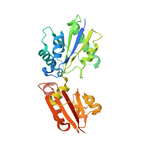 | |
UniProt | |||||
Find proteins for D5AV00 (Rhodobacter capsulatus (strain ATCC BAA-309 / NBRC 16581 / SB1003)) Explore D5AV00 Go to UniProtKB: D5AV00 | |||||
Entity Groups | |||||
| Sequence Clusters | 30% Identity50% Identity70% Identity90% Identity95% Identity100% Identity | ||||
| UniProt Group | D5AV00 | ||||
Sequence AnnotationsExpand | |||||
| |||||
| Ligands 2 Unique | |||||
|---|---|---|---|---|---|
| ID | Chains | Name / Formula / InChI Key | 2D Diagram | 3D Interactions | |
| SAH Query on SAH | C [auth A], F [auth B] | S-ADENOSYL-L-HOMOCYSTEINE C14 H20 N6 O5 S ZJUKTBDSGOFHSH-WFMPWKQPSA-N |  | ||
| ACT Query on ACT | D [auth A], E [auth A], G [auth B] | ACETATE ION C2 H3 O2 QTBSBXVTEAMEQO-UHFFFAOYSA-M |  | ||
| Length ( Å ) | Angle ( ˚ ) |
|---|---|
| a = 61.661 | α = 90 |
| b = 89.283 | β = 90 |
| c = 107.163 | γ = 90 |
| Software Name | Purpose |
|---|---|
| MOLREP | phasing |
| REFMAC | refinement |
| MOSFLM | data reduction |
| SCALA | data scaling |
RCSB PDB (citation) is hosted by
RCSB PDB is a member of the

