3N2C

Crystal structure of prolidase eah89906 complexed with n-methylphosphonate-l-proline


Experimental Data Snapshot

  • Method: X-RAY DIFFRACTION
  • Resolution: 2.81 Å
  • R-Value Free: 0.273 
  • R-Value Work: 0.220 
  • R-Value Observed: 0.222 

wwPDB Validation   3D Report Full Report


Ligand Structure Quality Assessment 


This is version 1.6 of the entry. See complete history


Literature

Functional identification and structure determination of two novel prolidases from cog1228 in the amidohydrolase superfamily .

Xiang, D.F.Patskovsky, Y.Xu, C.Fedorov, A.A.Fedorov, E.V.Sisco, A.A.Sauder, J.M.Burley, S.K.Almo, S.C.Raushel, F.M.

(2010) Biochemistry 49: 6791-6803

  • DOI: https://doi.org/10.1021/bi100897u
  • Primary Citation of Related Structures:  
    3FEQ, 3MKV, 3MTW, 3N2C

  • PubMed Abstract: 

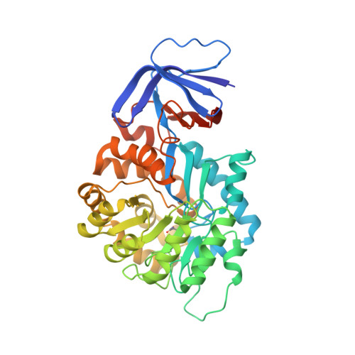
    Two uncharacterized enzymes from the amidohydrolase superfamily belonging to cog1228 were cloned, expressed, and purified to homogeneity. The two proteins, Sgx9260c ( gi|44242006 ) and Sgx9260b ( gi|44479596 ), were derived from environmental DNA samples originating from the Sargasso Sea. The catalytic function and substrate profiles for Sgx9260c and Sgx9260b were determined using a comprehensive library of dipeptides and N-acyl derivative of l-amino acids. Sgx9260c catalyzes the hydrolysis of Gly-l-Pro, l-Ala-l-Pro, and N-acyl derivatives of l-Pro. The best substrate identified to date is N-acetyl-l-Pro with a value of k(cat)/K(m) of 3 x 10(5) M(-1) s(-1). Sgx9260b catalyzes the hydrolysis of l-hydrophobic l-Pro dipeptides and N-acyl derivatives of l-Pro. The best substrate identified to date is N-propionyl-l-Pro with a value of k(cat)/K(m) of 1 x 10(5) M(-1) s(-1). Three-dimensional structures of both proteins were determined by X-ray diffraction methods (PDB codes 3MKV and 3FEQ ). These proteins fold as distorted (beta/alpha)(8)-barrels with two divalent cations in the active site. The structure of Sgx9260c was also determined as a complex with the N-methylphosphonate derivative of l-Pro (PDB code 3N2C ). In this structure the phosphonate moiety bridges the binuclear metal center, and one oxygen atom interacts with His-140. The alpha-carboxylate of the inhibitor interacts with Tyr-231. The proline side chain occupies a small substrate binding cavity formed by residues contributed from the loop that follows beta-strand 7 within the (beta/alpha)(8)-barrel. A total of 38 other proteins from cog1228 are predicted to have the same substrate profile based on conservation of the substrate binding residues. The structure of an evolutionarily related protein, Cc2672 from Caulobacter crecentus, was determined as a complex with the N-methylphosphonate derivative of l-arginine (PDB code 3MTW ).


  • Organizational Affiliation

    Department of Chemistry, P.O. Box 30012, Texas A&M University, College Station, Texas 77842-3012, USA.


Macromolecules
Find similar proteins by:  (by identity cutoff)  |  3D Structure
Entity ID: 1
MoleculeChains Sequence LengthOrganismDetailsImage
PROLIDASE
A, B, C, D, E
A, B, C, D, E, F, G, H, I, J, K, L, M, N, O, P
423unidentifiedMutation(s): 0 
UniProt
Find proteins for Q393A1 (Burkholderia lata (strain ATCC 17760 / DSM 23089 / LMG 22485 / NCIMB 9086 / R18194 / 383))
Explore Q393A1 
Go to UniProtKB:  Q393A1
Entity Groups  
Sequence Clusters30% Identity50% Identity70% Identity90% Identity95% Identity100% Identity
UniProt GroupQ393A1
Sequence Annotations
Expand
  • Reference Sequence
Small Molecules
Ligands 2 Unique
IDChains Name / Formula / InChI Key2D Diagram3D Interactions
LWY
Query on LWY

Download Ideal Coordinates CCD File 
BA [auth D]
CB [auth M]
EA [auth E]
FB [auth N]
HA [auth F]
BA [auth D],
CB [auth M],
EA [auth E],
FB [auth N],
HA [auth F],
IB [auth O],
KA [auth G],
LB [auth P],
NA [auth H],
QA [auth I],
S [auth A],
TA [auth J],
V [auth B],
WA [auth K],
Y [auth C],
ZA [auth L]
1-[(R)-hydroxy(methyl)phosphoryl]-L-proline
C6 H12 N O4 P
IIXOSEXCIQDQTO-YFKPBYRVSA-N
ZN
Query on ZN

Download Ideal Coordinates CCD File 
AA [auth D]
AB [auth M]
BB [auth M]
CA [auth E]
DA [auth E]
AA [auth D],
AB [auth M],
BB [auth M],
CA [auth E],
DA [auth E],
DB [auth N],
EB [auth N],
FA [auth F],
GA [auth F],
GB [auth O],
HB [auth O],
IA [auth G],
JA [auth G],
JB [auth P],
KB [auth P],
LA [auth H],
MA [auth H],
OA [auth I],
PA [auth I],
Q [auth A],
R [auth A],
RA [auth J],
SA [auth J],
T [auth B],
U [auth B],
UA [auth K],
VA [auth K],
W [auth C],
X [auth C],
XA [auth L],
YA [auth L],
Z [auth D]
ZINC ION
Zn
PTFCDOFLOPIGGS-UHFFFAOYSA-N
Modified Residues  1 Unique
IDChains TypeFormula2D DiagramParent
KCX
Query on KCX
A, B, C, D, E
A, B, C, D, E, F, G, H, I, J, K, L, M, N, O, P
L-PEPTIDE LINKINGC7 H14 N2 O4LYS
Binding Affinity Annotations 
IDSourceBinding Affinity
LWY PDBBind:  3N2C Ki: 7500 (nM) from 1 assay(s)
Experimental Data & Validation

Experimental Data

  • Method: X-RAY DIFFRACTION
  • Resolution: 2.81 Å
  • R-Value Free: 0.273 
  • R-Value Work: 0.220 
  • R-Value Observed: 0.222 
  • Space Group: P 1
Unit Cell:
Length ( Å )Angle ( ˚ )
a = 113.186α = 81.32
b = 108.035β = 80.47
c = 170.772γ = 73.76
Software Package:
Software NamePurpose
PHASERphasing
REFMACrefinement
HKL-2000data reduction
HKL-2000data scaling

Structure Validation

View Full Validation Report


Ligand Structure Quality Assessment 


Entry History 

Deposition Data

Revision History  (Full details and data files)

  • Version 1.0: 2010-06-02
    Type: Initial release
  • Version 1.1: 2011-07-13
    Changes: Version format compliance
  • Version 1.2: 2012-10-10
    Changes: Database references
  • Version 1.3: 2018-11-21
    Changes: Data collection, Structure summary
  • Version 1.4: 2021-02-10
    Changes: Database references, Derived calculations, Structure summary
  • Version 1.5: 2023-09-06
    Changes: Data collection, Database references, Refinement description
  • Version 1.6: 2023-11-22
    Changes: Data collection