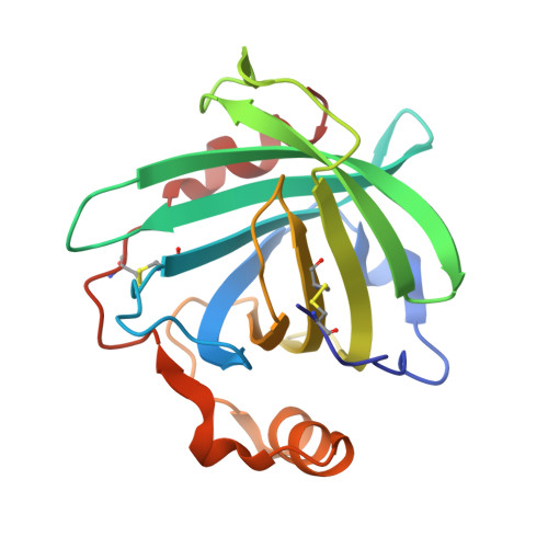
Formation of the complex of nitrite with the ferriheme b beta-barrel proteins nitrophorin 4 and nitrophorin 7.
He, C., Ogata, H., Knipp, M.(2010) Biochemistry 49: 5841-5851
- PubMed: 20524697
- DOI: https://doi.org/10.1021/bi100324z
- Primary Citation of Related Structures:
3MVF - PubMed Abstract:
The interaction of ferriheme proteins with nitrite has recently attracted interest as a source for NO or other nitrogen oxides in mammalian physiology. However, met-hemoglobin (metHb), which was suggested as a key player in this process, does not convert nitrite unless small amounts of NO are added in parallel. We have recently reported that, in contrast, nitrophorins (NPs) convert nitrite as the sole substrate to form NO even at pH 7.5, which is an unprecedented case among ferrihemes [He, C., and Knipp, M. (2009) J. Am. Chem. Soc. 131, 12042-12043]. NPs, which comprise a class of unique heme b proteins from the saliva of the blood-sucking insect Rhodnius prolixus, appear in a number of concomitant isoproteins. Herein, the first spectroscopic characterization of the initial complexes of the two isoproteins NP4 and NP7 with nitrite is presented and compared to the data reported for metHb and met-myoglobin (metMb). Because upon nitrite binding, NPs, in contrast to metHb and metMb, continue to react with nitrite, resonance Raman spectroscopy and continuous wave electron paramagnetic resonance spectroscopy were applied to frozen samples. As a result, the existence of two six-coordinate ferriheme low-spin complexes was established. Furthermore, X-ray crystallography of NP4 crystals soaked with nitrite revealed the formation of an eta(1)-N nitro complex, which is in contrast to the eta(1)-O-bound nitrite in metMb and metHb. Stopped-flow kinetic experiments show that although the ligand dissociation constants of NP4 and NP7 (15-190 M(-1)) are comparable to those of metHb and metMb, the rates of ligand binding and release are significantly slower. Moreover, not only the reaction kinetics but also electron paramagnetic resonance spectroscopy reveals notable differences between the two isoproteins.
Organizational Affiliation:
Max-Planck-Institut für Bioanorganische Chemie, Stiftstrasse 34-36, D-45470 Mülheim an der Ruhr, Germany.