3MIP

I-MsoI re-designed for altered DNA cleavage specificity (-8GCG)


Experimental Data Snapshot

  • Method: X-RAY DIFFRACTION
  • Resolution: 2.40 Å
  • R-Value Free: 0.285 
  • R-Value Work: 0.226 
  • R-Value Observed: 0.232 

wwPDB Validation   3D Report Full Report


This is version 1.2 of the entry. See complete history


Literature

Computational reprogramming of homing endonuclease specificity at multiple adjacent base pairs.

Ashworth, J.Taylor, G.K.Havranek, J.J.Quadri, S.A.Stoddard, B.L.Baker, D.

(2010) Nucleic Acids Res 38: 5601-5608

  • DOI: https://doi.org/10.1093/nar/gkq283
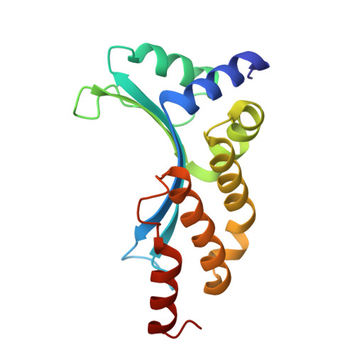
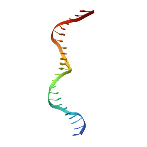
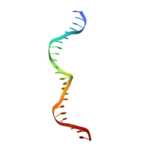
  • Primary Citation of Related Structures:  
    3KO2, 3MIP, 3MIS

  • PubMed Abstract: 

    Site-specific homing endonucleases are capable of inducing gene conversion via homologous recombination. Reprogramming their cleavage specificities allows the targeting of specific biological sites for gene correction or conversion. We used computational protein design to alter the cleavage specificity of I-MsoI for three contiguous base pair substitutions, resulting in an endonuclease whose activity and specificity for its new site rival that of wild-type I-MsoI for the original site. Concerted design for all simultaneous substitutions was more successful than a modular approach against individual substitutions, highlighting the importance of context-dependent redesign and optimization of protein-DNA interactions. We then used computational design based on the crystal structure of the designed complex, which revealed significant unanticipated shifts in DNA conformation, to create an endonuclease that specifically cleaves a site with four contiguous base pair substitutions. Our results demonstrate that specificity switches for multiple concerted base pair substitutions can be computationally designed, and that iteration between design and structure determination provides a route to large scale reprogramming of specificity.


  • Organizational Affiliation

    Department of Biochemistry, University of Washington, Seattle, WA 98195, USA. ashwortj@u.washington.edu


Macromolecules

Find similar proteins by:  (by identity cutoff)  |  3D Structure
Entity ID: 1
MoleculeChains Sequence LengthOrganismDetailsImage
Mso-8G
A, B
161synthetic constructMutation(s): 0 
Entity Groups  
Sequence Clusters30% Identity50% Identity70% Identity90% Identity95% Identity100% Identity
Sequence Annotations
Expand
  • Reference Sequence

Find similar nucleic acids by:  Sequence   |   3D Structure  

Entity ID: 2
MoleculeChains LengthOrganismImage
DNA (5'-D(*GP*CP*AP*GP*GP*CP*GP*GP*TP*CP*GP*TP*GP*AP*GP*AP*CP*CP*GP*CP*TP*CP*CP*G)-3')24N/A
Sequence Annotations
Expand
  • Reference Sequence

Find similar nucleic acids by:  Sequence   |   3D Structure  

Entity ID: 3
MoleculeChains LengthOrganismImage
DNA (5'-D(*CP*GP*GP*AP*GP*CP*GP*GP*TP*CP*TP*CP*AP*CP*GP*AP*CP*CP*GP*CP*CP*TP*GP*C)-3')24N/A
Sequence Annotations
Expand
  • Reference Sequence
Experimental Data & Validation

Experimental Data

  • Method: X-RAY DIFFRACTION
  • Resolution: 2.40 Å
  • R-Value Free: 0.285 
  • R-Value Work: 0.226 
  • R-Value Observed: 0.232 
  • Space Group: P 1
Unit Cell:
Length ( Å )Angle ( ˚ )
a = 42.539α = 96.45
b = 42.611β = 107.04
c = 71.524γ = 108.56
Software Package:
Software NamePurpose
CrystalCleardata collection
PHENIXmodel building
PHENIXrefinement
HKL-2000data reduction
HKL-2000data scaling
PHENIXphasing

Structure Validation

View Full Validation Report


Entry History 

Deposition Data

Revision History  (Full details and data files)

  • Version 1.0: 2010-05-19
    Type: Initial release
  • Version 1.1: 2011-07-13
    Changes: Version format compliance
  • Version 1.2: 2024-02-21
    Changes: Data collection, Database references, Derived calculations