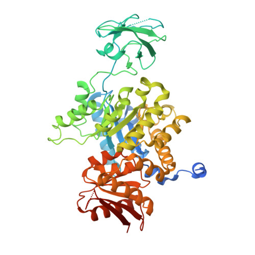
Crystal structure of CGD1_2040, a pyruvate kinase from cryptosporidium Parvum
Wernimont, A.K., Hutchinson, A., Hassanali, A., Kozieradzki, I., Cossar, D., Bochkarev, A., Arrowsmith, C.H., Edwards, A.M., Bountra, C., Weigelt, J., Hui, R., Hills, T., Pizarro, J.C.To be published.