3M22

Crystal structure of TagRFP fluorescent protein


Experimental Data Snapshot

  • Method: X-RAY DIFFRACTION
  • Resolution: 2.20 Å
  • R-Value Free: 0.179 
  • R-Value Work: 0.160 
  • R-Value Observed: 0.161 

wwPDB Validation   3D Report Full Report


This is version 1.3 of the entry. See complete history


Literature

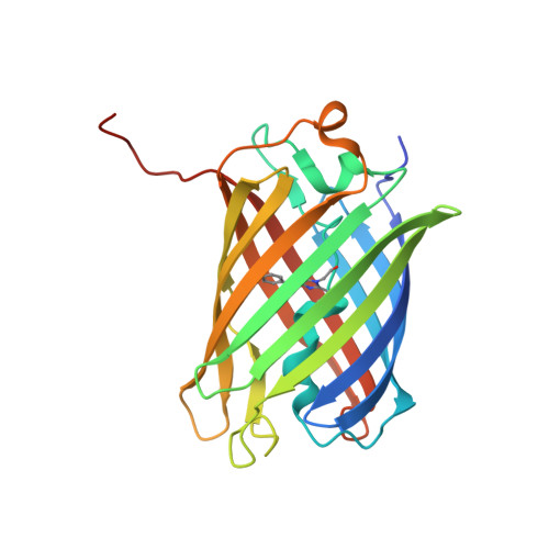
Structural characterization of acylimine-containing blue and red chromophores in mTagBFP and TagRFP fluorescent proteins.

Subach, O.M.Malashkevich, V.N.Zencheck, W.D.Morozova, K.S.Piatkevich, K.D.Almo, S.C.Verkhusha, V.V.

(2010) Chem Biol 17: 333-341

  • DOI: https://doi.org/10.1016/j.chembiol.2010.03.005
  • Primary Citation of Related Structures:  
    3M22, 3M24

  • PubMed Abstract: 

    We determined the 2.2 A crystal structures of the red fluorescent protein TagRFP and its derivative, the blue fluorescent protein mTagBFP. The crystallographic analysis is consistent with a model in which TagRFP has the trans coplanar anionic chromophore with the conjugated pi-electron system, similar to that of DsRed-like chromophores. Refined conformation of mTagBFP suggests the presence of an N-acylimine functionality in its chromophore and single C(alpha)-C(beta) bond in the Tyr64 side chain. Mass spectrum of mTagBFP chromophore-bearing peptide indicates a loss of 20 Da upon maturation, whereas tandem mass spectrometry reveals that the C(alpha)-N bond in Leu63 is oxidized. These data indicate that mTagBFP has a new type of the chromophore, N-[(5-hydroxy-1H-imidazole-2-yl)methylidene]acetamide. We propose a chemical mechanism in which the DsRed-like chromophore is formed via the mTagBFP-like blue intermediate.


  • Organizational Affiliation

    Department of Anatomy and Structural Biology and Gruss-Lipper Biophotonics Center, Albert Einstein College of Medicine, 1300 Morris Park Avenue, Bronx, NY10461, USA.


Macromolecules
Find similar proteins by:  (by identity cutoff)  |  3D Structure
Entity ID: 1
MoleculeChains Sequence LengthOrganismDetailsImage
TagRFP
A, B, C, D
235synthetic constructMutation(s): 0 
Entity Groups  
Sequence Clusters30% Identity50% Identity70% Identity90% Identity95% Identity100% Identity
Sequence Annotations
Expand
  • Reference Sequence
Small Molecules
Modified Residues  1 Unique
IDChains TypeFormula2D DiagramParent
NRQ
Query on NRQ
A, B, C, D
L-PEPTIDE LINKINGC16 H17 N3 O4 SMET, TYR, GLY
Experimental Data & Validation

Experimental Data

  • Method: X-RAY DIFFRACTION
  • Resolution: 2.20 Å
  • R-Value Free: 0.179 
  • R-Value Work: 0.160 
  • R-Value Observed: 0.161 
  • Space Group: I 41
Unit Cell:
Length ( Å )Angle ( ˚ )
a = 130.989α = 90
b = 130.989β = 90
c = 105.966γ = 90
Software Package:
Software NamePurpose
DENZOdata reduction
SCALEPACKdata scaling
REFMACrefinement
PDB_EXTRACTdata extraction
CBASSdata collection
HKL-2000data reduction
HKL-2000data scaling
PHASESphasing

Structure Validation

View Full Validation Report


Entry History 

Deposition Data

Revision History  (Full details and data files)

  • Version 1.0: 2010-05-12
    Type: Initial release
  • Version 1.1: 2011-07-13
    Changes: Version format compliance
  • Version 1.2: 2016-11-23
    Changes: Database references
  • Version 1.3: 2017-11-08
    Changes: Refinement description