3L3V

Structure of HIV-1 integrase core domain in complex with sucrose


Experimental Data Snapshot

  • Method: X-RAY DIFFRACTION
  • Resolution: 2.00 Å
  • R-Value Free: 0.244 
  • R-Value Work: 0.194 
  • R-Value Observed: 0.196 

wwPDB Validation   3D Report Full Report


This is version 2.2 of the entry. See complete history


Literature

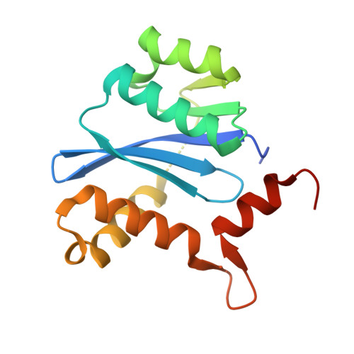
Crystal structure of the HIV-1 integrase core domain in complex with sucrose reveals details of an allosteric inhibitory binding site

Wielens, J.Headey, S.J.Jeevarajah, D.Rhodes, D.I.Deadman, J.Chalmers, D.K.Scanlon, M.J.Parker, M.W.

(2010) FEBS Lett 584: 1455-1462

  • DOI: https://doi.org/10.1016/j.febslet.2010.03.016
  • Primary Citation of Related Structures:  
    3L3U, 3L3V

  • PubMed Abstract: 

    HIV integrase (IN) is an essential enzyme in HIV replication and an important target for drug design. IN has been shown to interact with a number of cellular and viral proteins during the integration process. Disruption of these important interactions could provide a mechanism for allosteric inhibition of IN. We present the highest resolution crystal structure of the IN core domain to date. We also present a crystal structure of the IN core domain in complex with sucrose which is bound at the dimer interface in a region that has previously been reported to bind integrase inhibitors.


  • Organizational Affiliation

    Medicinal Chemistry and Drug Action, Monash Institute of Pharmaceutical Sciences, Parkville, Victoria, Australia.


Macromolecules
Find similar proteins by:  (by identity cutoff)  |  3D Structure
Entity ID: 1
MoleculeChains Sequence LengthOrganismDetailsImage
POL polyprotein
A, B
163Human immunodeficiency virus 1Mutation(s): 4 
Gene Names: pol
UniProt
Find proteins for Q72498 (Human immunodeficiency virus 1)
Explore Q72498 
Go to UniProtKB:  Q72498
Entity Groups  
Sequence Clusters30% Identity50% Identity70% Identity90% Identity95% Identity100% Identity
UniProt GroupQ72498
Sequence Annotations
Expand
  • Reference Sequence
Oligosaccharides

Help

Entity ID: 2
MoleculeChains Length2D Diagram Glycosylation3D Interactions
beta-D-fructofuranose-(2-1)-alpha-D-glucopyranose
C
2N/A
Glycosylation Resources
GlyTouCan:  G05551OP
GlyCosmos:  G05551OP
Biologically Interesting Molecules (External Reference) 1 Unique
Experimental Data & Validation

Experimental Data

  • Method: X-RAY DIFFRACTION
  • Resolution: 2.00 Å
  • R-Value Free: 0.244 
  • R-Value Work: 0.194 
  • R-Value Observed: 0.196 
  • Space Group: P 21 21 21
Unit Cell:
Length ( Å )Angle ( ˚ )
a = 60.085α = 90
b = 60.714β = 90
c = 81.463γ = 90
Software Package:
Software NamePurpose
MOSFLMdata reduction
SCALAdata scaling
AMoREphasing
REFMACrefinement
PDB_EXTRACTdata extraction
Blu-Icedata collection
PHENIXrefinement

Structure Validation

View Full Validation Report


Entry History 

Deposition Data

Revision History  (Full details and data files)

  • Version 1.0: 2010-03-31
    Type: Initial release
  • Version 1.1: 2011-07-13
    Changes: Version format compliance
  • Version 1.2: 2014-02-12
    Changes: Database references
  • Version 2.0: 2020-07-29
    Type: Remediation
    Reason: Carbohydrate remediation
    Changes: Atomic model, Data collection, Derived calculations, Non-polymer description, Structure summary
  • Version 2.1: 2021-11-10
    Changes: Database references, Structure summary
  • Version 2.2: 2023-11-01
    Changes: Data collection, Refinement description