3L2B

Crystal structure of the CBS and DRTGG domains of the regulatory region of Clostridium perfringens pyrophosphatase complexed with activator, diadenosine tetraphosphate


Experimental Data Snapshot

  • Method: X-RAY DIFFRACTION
  • Resolution: 2.27 Å
  • R-Value Free: 0.259 
  • R-Value Work: 0.209 
  • R-Value Observed: 0.211 

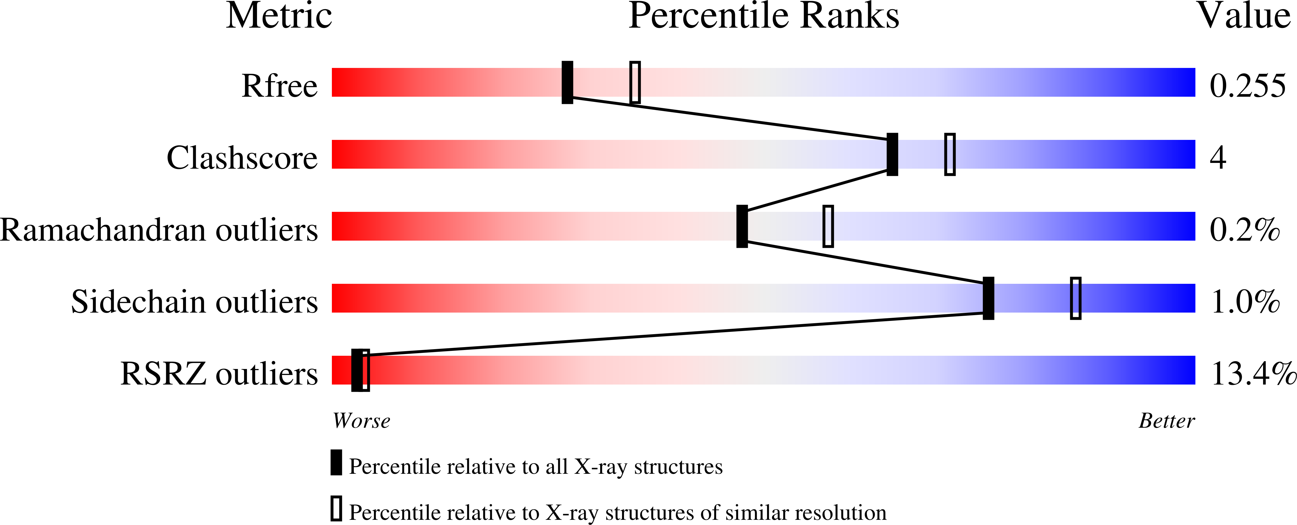
wwPDB Validation   3D Report Full Report


Ligand Structure Quality Assessment 


This is version 1.2 of the entry. See complete history


Literature

Crystal Structures of the CBS and DRTGG Domains of the Regulatory Region of Clostridiumperfringens Pyrophosphatase Complexed with the Inhibitor, AMP, and Activator, Diadenosine Tetraphosphate.

Tuominen, H.Salminen, A.Oksanen, E.Jamsen, J.Heikkila, O.Lehtio, L.Magretova, N.N.Goldman, A.Baykov, A.A.Lahti, R.

(2010) J Mol Biol 

  • DOI: https://doi.org/10.1016/j.jmb.2010.03.019
  • Primary Citation of Related Structures:  
    3L2B, 3L31

  • PubMed Abstract: 

    Nucleotide-binding cystathionine beta-synthase (CBS) domains serve as regulatory units in numerous proteins distributed in all kingdoms of life. However, the underlying regulatory mechanisms remain to be established. Recently, we described a subfamily of CBS domain-containing pyrophosphatases (PPases) within family II PPases. Here, we express a novel CBS-PPase from Clostridium perfringens (CPE2055) and show that the enzyme is inhibited by AMP and activated by a novel effector, diadenosine 5',5-P1,P4-tetraphosphate (AP(4)A). The structures of the AMP and AP(4)A complexes of the regulatory region of C. perfringens PPase (cpCBS), comprising a pair of CBS domains interlinked by a DRTGG domain, were determined at 2.3 A resolution using X-ray crystallography. The structures obtained are the first structures of a DRTGG domain as part of a larger protein structure. The AMP complex contains two AMP molecules per cpCBS dimer, each bound to a single monomer, whereas in the activator-bound complex, one AP(4)A molecule bridges two monomers. In the nucleotide-bound structures, activator binding induces significant opening of the CBS domain interface, compared with the inhibitor complex. These results provide structural insight into the mechanism of CBS-PPase regulation by nucleotides.


  • Organizational Affiliation

    Department of Biochemistry and Food Chemistry, University of Turku, Vatselankatu 2, FI-20014 Turku, Finland.


Macromolecules
Find similar proteins by:  (by identity cutoff)  |  3D Structure
Entity ID: 1
MoleculeChains Sequence LengthOrganismDetailsImage
Probable manganase-dependent inorganic pyrophosphatase
A, B
245Clostridium perfringens str. 13Mutation(s): 0 
Gene Names: CPE2055
EC: 3.6.1.1
UniProt
Find proteins for Q8XIQ9 (Clostridium perfringens (strain 13 / Type A))
Explore Q8XIQ9 
Go to UniProtKB:  Q8XIQ9
Entity Groups  
Sequence Clusters30% Identity50% Identity70% Identity90% Identity95% Identity100% Identity
UniProt GroupQ8XIQ9
Sequence Annotations
Expand
  • Reference Sequence
Small Molecules
Ligands 1 Unique
IDChains Name / Formula / InChI Key2D Diagram3D Interactions
B4P
Query on B4P

Download Ideal Coordinates CCD File 
C [auth A]BIS(ADENOSINE)-5'-TETRAPHOSPHATE
C20 H28 N10 O19 P4
YOAHKNVSNCMZGQ-XPWFQUROSA-N
Experimental Data & Validation

Experimental Data

  • Method: X-RAY DIFFRACTION
  • Resolution: 2.27 Å
  • R-Value Free: 0.259 
  • R-Value Work: 0.209 
  • R-Value Observed: 0.211 
  • Space Group: P 21 21 21
Unit Cell:
Length ( Å )Angle ( ˚ )
a = 63.847α = 90
b = 71.764β = 90
c = 116.334γ = 90
Software Package:
Software NamePurpose
XSCALEdata scaling
MOLREPphasing
REFMACrefinement
PDB_EXTRACTdata extraction
XDSdata scaling
XDSdata reduction

Structure Validation

View Full Validation Report


Ligand Structure Quality Assessment 


Entry History 

Deposition Data

Revision History  (Full details and data files)

  • Version 1.0: 2010-04-21
    Type: Initial release
  • Version 1.1: 2011-07-13
    Changes: Version format compliance
  • Version 1.2: 2023-11-01
    Changes: Data collection, Database references, Derived calculations, Refinement description