3KX2

Crystal structure of Prp43p in complex with ADP


Experimental Data Snapshot

  • Method: X-RAY DIFFRACTION
  • Resolution: 2.20 Å
  • R-Value Free: 0.250 
  • R-Value Work: 0.206 
  • R-Value Observed: 0.208 

wwPDB Validation   3D Report Full Report


Ligand Structure Quality Assessment 


This is version 1.3 of the entry. See complete history


Literature

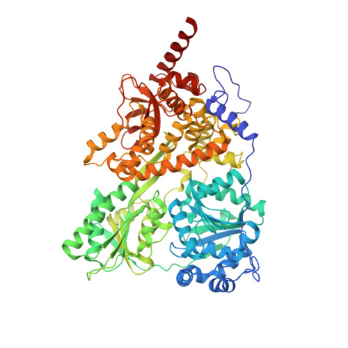
Structural basis for the function of DEAH helicases

He, Y.Andersen, G.R.Nielsen, K.H.

(2010) EMBO Rep 11: 180-186

  • DOI: https://doi.org/10.1038/embor.2010.11
  • Primary Citation of Related Structures:  
    3KX2

  • PubMed Abstract: 

    DEAH helicases participate in pre-messenger RNA splicing and ribosome biogenesis. The structure of yeast Prp43p-ADP reveals the homology of DEAH helicases to DNA helicases and the presence of an oligonucleotide-binding motif. A beta-hairpin from the second RecA domain is wedged between two carboxy-terminal domains and blocks access to the occluded RNA binding site formed by the RecA domains and a C-terminal domain. ATP binding and hydrolysis are likely to induce conformational changes in the hairpin that are important for RNA unwinding or ribonucleoprotein remodelling. The structure of Prp43p provides the framework for functional and genetic analysis of all DEAH helicases.


  • Organizational Affiliation

    Department of Molecular Biology, Aarhus University, Aarhus, Denmark.


Macromolecules
Find similar proteins by:  (by identity cutoff)  |  3D Structure
Entity ID: 1
MoleculeChains Sequence LengthOrganismDetailsImage
Pre-mRNA-splicing factor ATP-dependent RNA helicase PRP43A [auth B],
B [auth A]
767Saccharomyces cerevisiae S288CMutation(s): 0 
Gene Names: YGL120C
EC: 3.6.1
UniProt
Find proteins for P53131 (Saccharomyces cerevisiae (strain ATCC 204508 / S288c))
Explore P53131 
Go to UniProtKB:  P53131
Entity Groups  
Sequence Clusters30% Identity50% Identity70% Identity90% Identity95% Identity100% Identity
UniProt GroupP53131
Sequence Annotations
Expand
  • Reference Sequence
Experimental Data & Validation

Experimental Data

  • Method: X-RAY DIFFRACTION
  • Resolution: 2.20 Å
  • R-Value Free: 0.250 
  • R-Value Work: 0.206 
  • R-Value Observed: 0.208 
  • Space Group: P 32 2 1
Unit Cell:
Length ( Å )Angle ( ˚ )
a = 118.152α = 90
b = 118.152β = 90
c = 253.601γ = 120
Software Package:
Software NamePurpose
SHELXSphasing
PHENIXrefinement
XDSdata reduction
XSCALEdata scaling

Structure Validation

View Full Validation Report


Ligand Structure Quality Assessment 


Entry History 

Deposition Data

Revision History  (Full details and data files)

  • Version 1.0: 2010-01-26
    Type: Initial release
  • Version 1.1: 2011-07-13
    Changes: Version format compliance
  • Version 1.2: 2017-11-01
    Changes: Data collection, Refinement description
  • Version 1.3: 2024-03-20
    Changes: Data collection, Database references, Derived calculations