Human cytochrome P450 2E1 structures with fatty acid analogs reveal a previously unobserved binding mode.
Porubsky, P.R., Battaile, K.P., Scott, E.E.(2010) J Biol Chem 285: 22282-22290
- PubMed: 20463018
- DOI: https://doi.org/10.1074/jbc.M110.109017
- Primary Citation of Related Structures:
3GPH, 3KOH, 3LC4 - PubMed Abstract:
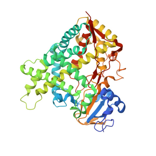
Human microsomal cytochrome P450 (CYP) 2E1 is widely known for its ability to oxidize >70 different, mostly compact, low molecular weight drugs and other xenobiotic compounds. In addition CYP2E1 oxidizes much larger C9-C20 fatty acids that can serve as endogenous signaling molecules. Previously structures of CYP2E1 with small molecules revealed a small, compact CYP2E1 active site, which would be insufficient to accommodate medium and long chain fatty acids without conformational changes in the protein. In the current work we have determined how CYP2E1 can accommodate a series of fatty acid analogs by cocrystallizing CYP2E1 with omega-imidazolyl-octanoic fatty acid, omega-imidazolyl-decanoic fatty acid, and omega-imidazolyl-dodecanoic fatty acid. In each structure direct coordination of the imidazole nitrogen to the heme iron mimics the position required for native fatty acid substrates to yield the omega-1 hydroxylated metabolites that predominate experimentally. In each case rotation of a single Phe(298) side chain merges the active site with an adjacent void, significantly altering the active site size and topology to accommodate fatty acids. The binding of these fatty acid ligands is directly opposite the channel to the protein surface and the binding observed for fatty acids in the bacterial cytochrome P450 BM3 (CYP102A1) from Bacillus megaterium. Instead of the BM3-like binding mode in the CYP2E1 channel, these structures reveal interactions between the fatty acid carboxylates and several residues in the F, G, and B' helices at successive distances from the active site.
Organizational Affiliation:
Department of Medicinal Chemistry, The University of Kansas, Lawrence, Kansas 66045, USA.