3KMV

Crystal structure of CBM42A from Clostridium thermocellum


Experimental Data Snapshot

  • Method: X-RAY DIFFRACTION
  • Resolution: 1.80 Å
  • R-Value Free: 0.192 
  • R-Value Work: 0.158 
  • R-Value Observed: 0.160 

wwPDB Validation   3D Report Full Report


This is version 1.3 of the entry. See complete history


Literature

Family 42 carbohydrate-binding modules display multiple arabinoxylan-binding interfaces presenting different ligand affinities.

Ribeiro, T.Santos-Silva, T.Alves, V.D.Dias, F.M.Luis, A.S.Prates, J.A.Ferreira, L.M.Romao, M.J.Fontes, C.M.

(2010) Biochim Biophys Acta 1804: 2054-2062

  • DOI: https://doi.org/10.1016/j.bbapap.2010.07.006
  • Primary Citation of Related Structures:  
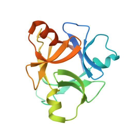
    3KMV

  • PubMed Abstract: 

    Enzymes that degrade plant cell wall polysaccharides display a modular architecture comprising a catalytic domain bound to one or more non-catalytic carbohydrate-binding modules (CBMs). CBMs display considerable variation in primary structure and are grouped into 59 sequence-based families organized in the Carbohydrate-Active enZYme (CAZy) database. Here we report the crystal structure of CtCBM42A together with the biochemical characterization of two other members of family 42 CBMs from Clostridium thermocellum. CtCBM42A, CtCBM42B and CtCBM42C bind specifically to the arabinose side-chains of arabinoxylans and arabinan, suggesting that various cellulosomal components are targeted to these regions of the plant cell wall. The structure of CtCBM42A displays a beta-trefoil fold, which comprises 3 sub-domains designated as alpha, beta and gamma. Each one of the three sub-domains presents a putative carbohydrate-binding pocket where an aspartate residue located in a central position dominates ligand recognition. Intriguingly, the gamma sub-domain of CtCBM42A is pivotal for arabinoxylan binding, while the concerted action of beta and gamma sub-domains of CtCBM42B and CtCBM42C is apparently required for ligand sequestration. Thus, this work reveals that the binding mechanism of CBM42 members is in contrast with that of homologous CBM13s where recognition of complex polysaccharides results from the cooperative action of three protein sub-domains presenting similar affinities.


  • Organizational Affiliation

    CIISA-Faculdade de Medicina Veterinária, Pólo Universitário do Alto da Ajuda, Avenida da Universidade Técnica, 1300-477 Lisboa, Portugal.


Macromolecules
Find similar proteins by:  (by identity cutoff)  |  3D Structure
Entity ID: 1
MoleculeChains Sequence LengthOrganismDetailsImage
Alpha-L-arabinofuranosidase B
A, B, C, D, E
A, B, C, D, E, F, G, H
157Acetivibrio thermocellus ATCC 27405Mutation(s): 0 
Gene Names: Cthe_0015
UniProt
Find proteins for A3DBC8 (Acetivibrio thermocellus (strain ATCC 27405 / DSM 1237 / JCM 9322 / NBRC 103400 / NCIMB 10682 / NRRL B-4536 / VPI 7372))
Explore A3DBC8 
Go to UniProtKB:  A3DBC8
Entity Groups  
Sequence Clusters30% Identity50% Identity70% Identity90% Identity95% Identity100% Identity
UniProt GroupA3DBC8
Sequence Annotations
Expand
  • Reference Sequence
Small Molecules
Ligands 4 Unique
IDChains Name / Formula / InChI Key2D Diagram3D Interactions
GOL
Query on GOL

Download Ideal Coordinates CCD File 
BA [auth E]
FA [auth F]
I [auth A]
NA [auth H]
T [auth C]
BA [auth E],
FA [auth F],
I [auth A],
NA [auth H],
T [auth C],
Z [auth D]
GLYCEROL
C3 H8 O3
PEDCQBHIVMGVHV-UHFFFAOYSA-N
ACT
Query on ACT

Download Ideal Coordinates CCD File 
IA [auth G]ACETATE ION
C2 H3 O2
QTBSBXVTEAMEQO-UHFFFAOYSA-M
FMT
Query on FMT

Download Ideal Coordinates CCD File 
CA [auth E]
DA [auth E]
GA [auth F]
J [auth A]
JA [auth G]
CA [auth E],
DA [auth E],
GA [auth F],
J [auth A],
JA [auth G],
K [auth A],
KA [auth G],
L [auth A],
LA [auth G],
M [auth A],
O [auth B],
OA [auth H],
P [auth B],
Q [auth B],
R [auth B],
U [auth C],
V [auth C],
W [auth C],
X [auth C]
FORMIC ACID
C H2 O2
BDAGIHXWWSANSR-UHFFFAOYSA-N
CA
Query on CA

Download Ideal Coordinates CCD File 
AA [auth D]
EA [auth E]
HA [auth F]
MA [auth G]
N [auth A]
AA [auth D],
EA [auth E],
HA [auth F],
MA [auth G],
N [auth A],
PA [auth H],
S [auth B],
Y [auth C]
CALCIUM ION
Ca
BHPQYMZQTOCNFJ-UHFFFAOYSA-N
Experimental Data & Validation

Experimental Data

  • Method: X-RAY DIFFRACTION
  • Resolution: 1.80 Å
  • R-Value Free: 0.192 
  • R-Value Work: 0.158 
  • R-Value Observed: 0.160 
  • Space Group: P 32 2 1
Unit Cell:
Length ( Å )Angle ( ˚ )
a = 106.37α = 90
b = 106.37β = 90
c = 237.564γ = 120
Software Package:
Software NamePurpose
MOSFLMdata reduction
SCALAdata scaling
PHASERphasing
REFMACrefinement
PDB_EXTRACTdata extraction
ADSCdata collection

Structure Validation

View Full Validation Report


Entry History 

Deposition Data

Revision History  (Full details and data files)

  • Version 1.0: 2010-08-04
    Type: Initial release
  • Version 1.1: 2011-07-13
    Changes: Source and taxonomy, Version format compliance
  • Version 1.2: 2018-01-24
    Changes: Structure summary
  • Version 1.3: 2023-09-06
    Changes: Data collection, Database references, Derived calculations, Refinement description