3KF3

Structure of fructofuranosidase from Schwanniomyces occidentalis complexed with fructose


Experimental Data Snapshot

  • Method: X-RAY DIFFRACTION
  • Resolution: 1.90 Å
  • R-Value Free: 0.261 
  • R-Value Work: 0.220 
  • R-Value Observed: 0.222 

wwPDB Validation   3D Report Full Report


Ligand Structure Quality Assessment 


This is version 1.3 of the entry. See complete history


Literature

Structural and kinetic analysis of Schwanniomyces occidentalis invertase reveals a new oligomerization pattern and the role of its supplementary domain in substrate binding

Alvaro-Benito, M.Polo, A.Gonzalez, B.Fernandez-Lobato, M.Sanz-Aparicio, J.

(2010) J Biol Chem 285: 13930-13941

  • DOI: https://doi.org/10.1074/jbc.M109.095430
  • Primary Citation of Related Structures:  
    3KF3, 3KF5

  • PubMed Abstract: 

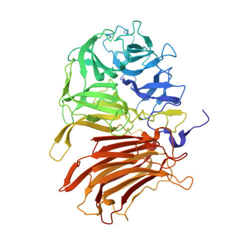
    Schwanniomyces occidentalis invertase is an extracellular enzyme that hydrolyzes sucrose and releases beta-fructose from various oligosaccharides and essential storage fructan polymers such as inulin. We report here the three-dimensional structure of Sw. occidentalis invertase at 2.9 A resolution and its complex with fructose at 1.9 A resolution. The monomer presents a bimodular arrangement common to other GH32 enzymes, with an N-terminal 5-fold beta-propeller catalytic domain and a C-terminal beta-sandwich domain for which the function has been unknown until now. However, the dimeric nature of Sw. occidentalis invertase reveals a unique active site cleft shaped by both subunits that may be representative of other yeast enzymes reported to be multimeric. Binding of the tetrasaccharide nystose and the polymer inulin was explored by docking analysis, which suggested that medium size and long substrates are recognized by residues from both subunits. The identified residues were mutated, and the enzymatic activity of the mutants against sucrose, nystose, and inulin were investigated by kinetic analysis. The replacements that showed the largest effect on catalytic efficiency were Q228V, a residue putatively involved in nystose and inulin binding, and S281I, involved in a polar link at the dimer interface. Moreover, a significant decrease in catalytic efficiency against inulin was observed in the mutants Q435A and Y462A, both located in the beta-sandwich domain of the second monomer. This highlights the essential function that oligomerization plays in substrate specificity and assigns, for the first time, a direct catalytic role to the supplementary domain of a GH32 enzyme.


  • Organizational Affiliation

    Centro de Biología Molecular Severo Ochoa, Departamento de Biología Molecular, Consejo Superior de Investigaciones Cientificas-Universidad Autónoma de Madrid, Cantoblanco, 28049 Madrid, Spain.


Macromolecules
Find similar proteins by:  (by identity cutoff)  |  3D Structure
Entity ID: 1
MoleculeChains Sequence LengthOrganismDetailsImage
Invertase
A, B
509Schwanniomyces occidentalisMutation(s): 0 
EC: 3.2.1.26
UniProt
Find proteins for P24133 (Schwanniomyces occidentalis)
Explore P24133 
Go to UniProtKB:  P24133
Entity Groups  
Sequence Clusters30% Identity50% Identity70% Identity90% Identity95% Identity100% Identity
UniProt GroupP24133
Sequence Annotations
Expand
  • Reference Sequence
Small Molecules
Ligands 2 Unique
IDChains Name / Formula / InChI Key2D Diagram3D Interactions
NAG
Query on NAG

Download Ideal Coordinates CCD File 
D [auth A]
E [auth A]
F [auth A]
G [auth A]
I [auth B]
D [auth A],
E [auth A],
F [auth A],
G [auth A],
I [auth B],
J [auth B],
K [auth B]
2-acetamido-2-deoxy-beta-D-glucopyranose
C8 H15 N O6
OVRNDRQMDRJTHS-FMDGEEDCSA-N
FRU
Query on FRU

Download Ideal Coordinates CCD File 
C [auth A],
H [auth B]
beta-D-fructofuranose
C6 H12 O6
RFSUNEUAIZKAJO-ARQDHWQXSA-N
Experimental Data & Validation

Experimental Data

  • Method: X-RAY DIFFRACTION
  • Resolution: 1.90 Å
  • R-Value Free: 0.261 
  • R-Value Work: 0.220 
  • R-Value Observed: 0.222 
  • Space Group: P 1 21 1
Unit Cell:
Length ( Å )Angle ( ˚ )
a = 60.87α = 90
b = 92.27β = 104.81
c = 116.28γ = 90
Software Package:
Software NamePurpose
SCALAdata scaling
MOLREPphasing
REFMACrefinement
PDB_EXTRACTdata extraction
ADSCdata collection
MOSFLMdata reduction

Structure Validation

View Full Validation Report


Ligand Structure Quality Assessment 


Entry History 

Deposition Data

Revision History  (Full details and data files)

  • Version 1.0: 2010-02-23
    Type: Initial release
  • Version 1.1: 2011-07-13
    Changes: Advisory, Refinement description, Version format compliance
  • Version 1.2: 2020-07-29
    Type: Remediation
    Reason: Carbohydrate remediation
    Changes: Data collection, Database references, Derived calculations, Structure summary
  • Version 1.3: 2023-11-01
    Changes: Data collection, Database references, Refinement description, Structure summary