3K40

Crystal structure of Drosophila 3,4-dihydroxyphenylalanine decarboxylase


Experimental Data Snapshot

  • Method: X-RAY DIFFRACTION
  • Resolution: 1.75 Å
  • R-Value Free: 0.226 
  • R-Value Work: 0.197 
  • R-Value Observed: 0.198 

wwPDB Validation   3D Report Full Report


This is version 1.3 of the entry. See complete history


Literature

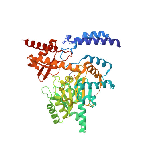
Crystal structure and substrate specificity of Drosophila 3,4-dihydroxyphenylalanine decarboxylase

Han, Q.Ding, H.Robinson, H.Christensen, B.M.Li, J.

(2010) PLoS One 5: e8826-e8826

  • DOI: https://doi.org/10.1371/journal.pone.0008826
  • Primary Citation of Related Structures:  
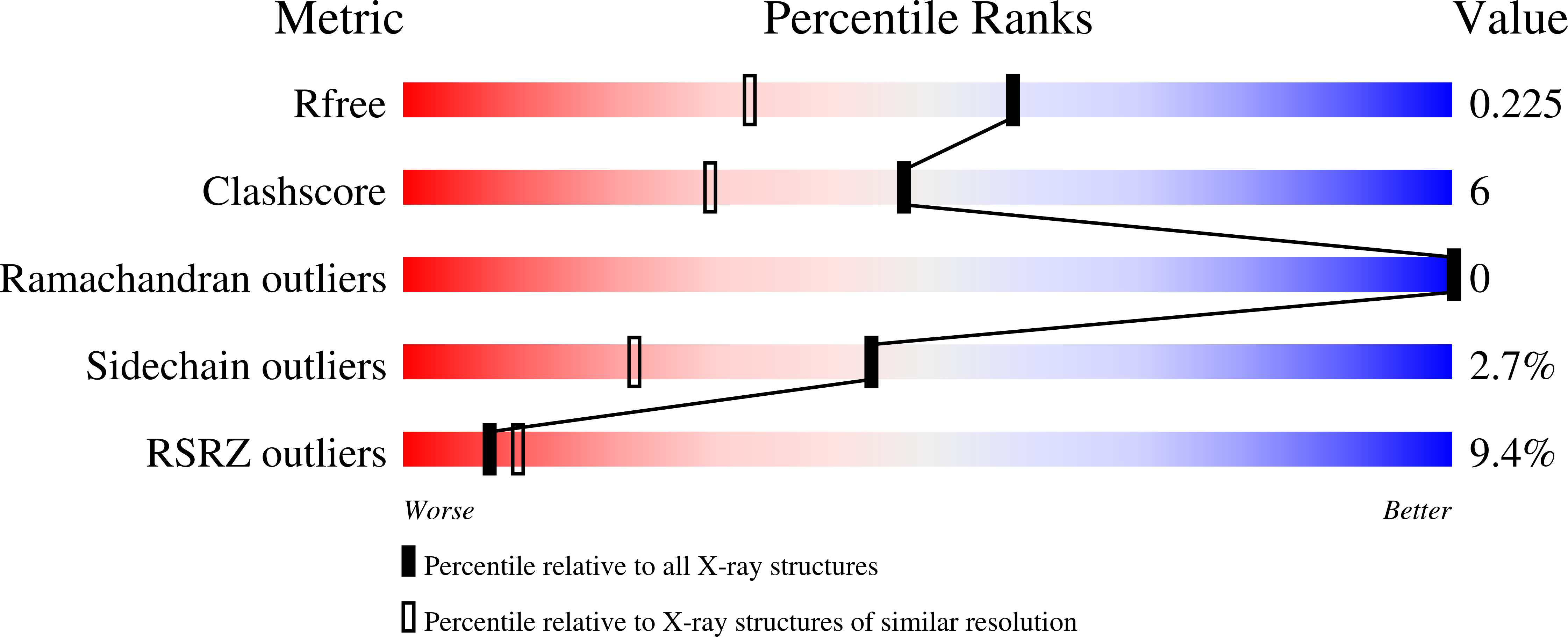
    3K40

  • PubMed Abstract: 

    3,4-Dihydroxyphenylalanine decarboxylase (DDC), also known as aromatic L-amino acid decarboxylase, catalyzes the decarboxylation of a number of aromatic L-amino acids. Physiologically, DDC is responsible for the production of dopamine and serotonin through the decarboxylation of 3,4-dihydroxyphenylalanine and 5-hydroxytryptophan, respectively. In insects, both dopamine and serotonin serve as classical neurotransmitters, neuromodulators, or neurohormones, and dopamine is also involved in insect cuticle formation, eggshell hardening, and immune responses. In this study, we expressed a typical DDC enzyme from Drosophila melanogaster, critically analyzed its substrate specificity and biochemical properties, determined its crystal structure at 1.75 Angstrom resolution, and evaluated the roles residues T82 and H192 play in substrate binding and enzyme catalysis through site-directed mutagenesis of the enzyme. Our results establish that this DDC functions exclusively on the production of dopamine and serotonin, with no activity to tyrosine or tryptophan and catalyzes the formation of serotonin more efficiently than dopamine. The crystal structure of Drosophila DDC and the site-directed mutagenesis study of the enzyme demonstrate that T82 is involved in substrate binding and that H192 is used not only for substrate interaction, but for cofactor binding of drDDC as well. Through comparative analysis, the results also provide insight into the structure-function relationship of other insect DDC-like proteins.


  • Organizational Affiliation

    Department of Biochemistry, Virginia Tech, Blacksburg, Virginia, United States of America.


Macromolecules
Find similar proteins by:  (by identity cutoff)  |  3D Structure
Entity ID: 1
MoleculeChains Sequence LengthOrganismDetailsImage
Aromatic-L-amino-acid decarboxylase
A, B
475Drosophila melanogasterMutation(s): 0 
Gene Names: CG10697Ddc
EC: 4.1.1.28
UniProt
Find proteins for P05031 (Drosophila melanogaster)
Explore P05031 
Go to UniProtKB:  P05031
Entity Groups  
Sequence Clusters30% Identity50% Identity70% Identity90% Identity95% Identity100% Identity
UniProt GroupP05031
Sequence Annotations
Expand
  • Reference Sequence
Small Molecules
Ligands 1 Unique
IDChains Name / Formula / InChI Key2D Diagram3D Interactions
GOL
Query on GOL

Download Ideal Coordinates CCD File 
C [auth A]GLYCEROL
C3 H8 O3
PEDCQBHIVMGVHV-UHFFFAOYSA-N
Modified Residues  1 Unique
IDChains TypeFormula2D DiagramParent
LLP
Query on LLP
A, B
L-PEPTIDE LINKINGC14 H22 N3 O7 PLYS
Experimental Data & Validation

Experimental Data

  • Method: X-RAY DIFFRACTION
  • Resolution: 1.75 Å
  • R-Value Free: 0.226 
  • R-Value Work: 0.197 
  • R-Value Observed: 0.198 
  • Space Group: P 21 21 2
Unit Cell:
Length ( Å )Angle ( ˚ )
a = 105.809α = 90
b = 108.597β = 90
c = 86.294γ = 90
Software Package:
Software NamePurpose
HKL-2000data collection
MOLREPphasing
REFMACrefinement
HKL-2000data reduction
HKL-2000data scaling

Structure Validation

View Full Validation Report


Entry History 

Deposition Data

Revision History  (Full details and data files)

  • Version 1.0: 2010-02-02
    Type: Initial release
  • Version 1.1: 2011-07-13
    Changes: Version format compliance
  • Version 1.2: 2023-09-06
    Changes: Data collection, Database references, Derived calculations, Refinement description
  • Version 1.3: 2023-11-22
    Changes: Data collection