3K1A

Insights into substrate binding at FeMo-cofactor in nitrogenase from the structure of an alpha-70Ile MoFe protein variant


Experimental Data Snapshot

  • Method: X-RAY DIFFRACTION
  • Resolution: 2.23 Å
  • R-Value Free: 0.254 
  • R-Value Work: 0.204 
  • R-Value Observed: 0.207 

wwPDB Validation   3D Report Full Report


Ligand Structure Quality Assessment 


This is version 1.4 of the entry. See complete history


Literature

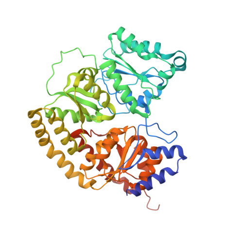
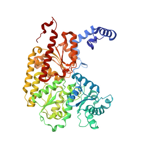
Insights into substrate binding at FeMo-cofactor in nitrogenase from the structure of an alpha-70(Ile) MoFe protein variant

Sarma, R.Barney, B.M.Keable, S.Dean, D.R.Seefeldt, L.C.Peters, J.W.

(2010) J Inorg Biochem 104: 385-389

  • DOI: https://doi.org/10.1016/j.jinorgbio.2009.11.009
  • Primary Citation of Related Structures:  
    3K1A

  • PubMed Abstract: 

    The X-ray crystal structure is presented for a nitrogenase MoFe protein where the alpha subunit residue at position 70 (alpha-70(Val)) has been substituted by the amino acid isoleucine (alpha-70(Ile)). Substitution of alpha-70(Val) by alpha-70(Ile) results in a MoFe protein that is hampered in its ability to reduce a range of substrates including acetylene and N(2), yet retains normal proton reduction activity. The 2.3A structure of the alpha-70(Ile) MoFe protein is compared to the alpha-70(Val) wild-type MoFe protein, revealing that the delta methyl group of alpha-70(Val) is positioned over Fe6 within the active site FeMo-cofactor. This work provides strong crystallographic support for the previously proposed model that substrates bind and are reduced at a single 4Fe-4S face of the FeMo-cofactor and that when alpha-70(Val) is substituted by alpha-70(Ile) access of substrates to Fe6 of this face is effectively blocked. Furthermore the detailed examination of the structure provides the basis for understanding the ability to trap and characterize hydrides in the variant, contributing significantly to our understanding of substrate access and substrate reduction at the FeMo-cofactor active site of nitrogenase.


  • Organizational Affiliation

    Department of Chemistry and Biochemistry and the Astrobiology Biogeocatalysis Research Center, Montana State University, Bozeman, MT 59717, USA.


Macromolecules
Find similar proteins by:  (by identity cutoff)  |  3D Structure
Entity ID: 1
MoleculeChains Sequence LengthOrganismDetailsImage
Nitrogenase molybdenum-iron protein alpha chain
A, C
491Azotobacter vinelandiiMutation(s): 1 
Gene Names: nifDKnifK
EC: 1.18.6.1
UniProt
Find proteins for P07328 (Azotobacter vinelandii)
Explore P07328 
Go to UniProtKB:  P07328
Entity Groups  
Sequence Clusters30% Identity50% Identity70% Identity90% Identity95% Identity100% Identity
UniProt GroupP07328
Sequence Annotations
Expand
  • Reference Sequence
Find similar proteins by:  (by identity cutoff)  |  3D Structure
Entity ID: 2
MoleculeChains Sequence LengthOrganismDetailsImage
Nitrogenase molybdenum-iron protein beta chain
B, D
522Azotobacter vinelandiiMutation(s): 0 
Gene Names: nifD
EC: 1.18.6.1
UniProt
Find proteins for P07329 (Azotobacter vinelandii)
Explore P07329 
Go to UniProtKB:  P07329
Entity Groups  
Sequence Clusters30% Identity50% Identity70% Identity90% Identity95% Identity100% Identity
UniProt GroupP07329
Sequence Annotations
Expand
  • Reference Sequence
Small Molecules
Ligands 4 Unique
IDChains Name / Formula / InChI Key2D Diagram3D Interactions
CFN
Query on CFN

Download Ideal Coordinates CCD File 
F [auth A],
I [auth C]
FE(7)-MO-S(9)-N CLUSTER
Fe7 Mo N S9
OSSJGSCTDHHOOW-UHFFFAOYSA-N
CLF
Query on CLF

Download Ideal Coordinates CCD File 
G [auth B],
L [auth D]
FE(8)-S(7) CLUSTER
Fe8 S7
JKVMXLBGZBULKV-UHFFFAOYSA-N
HCA
Query on HCA

Download Ideal Coordinates CCD File 
E [auth A],
J [auth C]
3-HYDROXY-3-CARBOXY-ADIPIC ACID
C7 H10 O7
XKJVEVRQMLKSMO-SSDOTTSWSA-N
CA
Query on CA

Download Ideal Coordinates CCD File 
H [auth B],
K [auth D]
CALCIUM ION
Ca
BHPQYMZQTOCNFJ-UHFFFAOYSA-N
Experimental Data & Validation

Experimental Data

  • Method: X-RAY DIFFRACTION
  • Resolution: 2.23 Å
  • R-Value Free: 0.254 
  • R-Value Work: 0.204 
  • R-Value Observed: 0.207 
  • Space Group: P 1 21 1
Unit Cell:
Length ( Å )Angle ( ˚ )
a = 77.019α = 90
b = 129.458β = 109.01
c = 107.088γ = 90
Software Package:
Software NamePurpose
HKL-2000data collection
PHASERphasing
REFMACrefinement
DENZOdata reduction
SCALEPACKdata scaling

Structure Validation

View Full Validation Report


Ligand Structure Quality Assessment 


Entry History 

Deposition Data

Revision History  (Full details and data files)

  • Version 1.0: 2010-02-16
    Type: Initial release
  • Version 1.1: 2011-07-13
    Changes: Version format compliance
  • Version 1.2: 2017-11-01
    Changes: Refinement description
  • Version 1.3: 2021-10-13
    Changes: Database references, Derived calculations
  • Version 1.4: 2023-09-06
    Changes: Data collection, Refinement description