3JR3

Sir2 bound to acetylated peptide


Experimental Data Snapshot

  • Method: X-RAY DIFFRACTION
  • Resolution: 1.50 Å
  • R-Value Free: 0.228 
  • R-Value Work: 0.205 
  • R-Value Observed: 0.206 

wwPDB Validation   3D Report Full Report


This is version 1.5 of the entry. See complete history


Literature

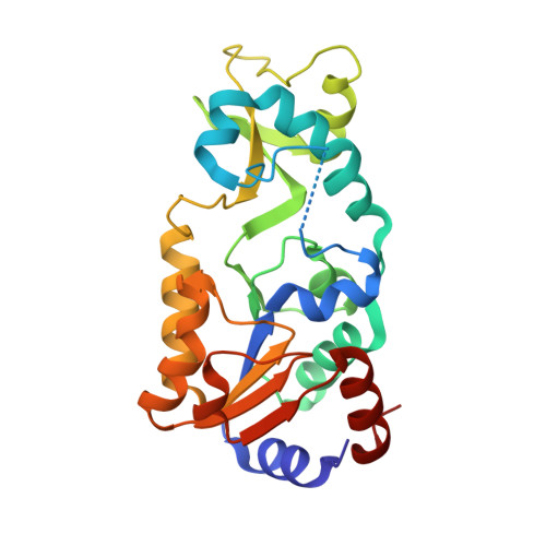
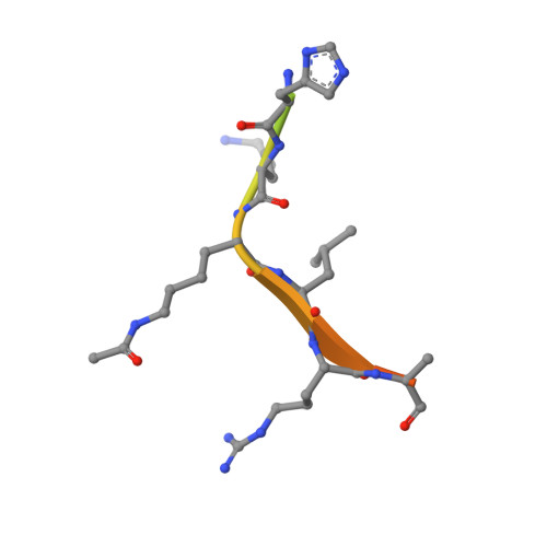
Structure-based mechanism of ADP-ribosylation by sirtuins.

Hawse, W.F.Wolberger, C.

(2009) J Biol Chem 284: 33654-33661

  • DOI: https://doi.org/10.1074/jbc.M109.024521
  • Primary Citation of Related Structures:  
    3JR3

  • PubMed Abstract: 

    Sirtuins comprise a family of enzymes found in all organisms, where they play a role in diverse processes including transcriptional silencing, aging, regulation of transcription, and metabolism. The predominant reaction catalyzed by these enzymes is NAD(+)-dependent lysine deacetylation, although some sirtuins exhibit a weaker ADP-ribosyltransferase activity. Although the Sir2 deacetylation mechanism is well established, much less is known about the Sir2 ADP-ribosylation reaction. We have studied the ADP-ribosylation activity of a bacterial sirtuin, Sir2Tm, and show that acetylated peptides containing arginine or lysine 2 residues C-terminal to the acetyl lysine, the +2 position, are preferentially ADP-ribosylated at the +2 residue. A structure of Sir2Tm bound to the acetylated +2 arginine peptide shows how this arginine could enter the active site and react with a deacetylation reaction intermediate to yield an ADP-ribosylated peptide. The new biochemical and structural studies presented here provide mechanistic insights into the Sir2 ADP-ribosylation reaction and will aid in identifying substrates of this reaction.


  • Organizational Affiliation

    Department of Biophysics and Biophysical Chemistry, The Johns Hopkins University School of Medicine, Baltimore, Maryland 21205, USA.


Macromolecules
Find similar proteins by:  (by identity cutoff)  |  3D Structure
Entity ID: 1
MoleculeChains Sequence LengthOrganismDetailsImage
NAD-dependent deacetylase246Thermotoga maritimaMutation(s): 1 
Gene Names: npdATM_0490
EC: 3.5.1
UniProt
Find proteins for Q9WYW0 (Thermotoga maritima (strain ATCC 43589 / DSM 3109 / JCM 10099 / NBRC 100826 / MSB8))
Explore Q9WYW0 
Go to UniProtKB:  Q9WYW0
Entity Groups  
Sequence Clusters30% Identity50% Identity70% Identity90% Identity95% Identity100% Identity
UniProt GroupQ9WYW0
Sequence Annotations
Expand
  • Reference Sequence

Find similar proteins by:  Sequence   |   3D Structure  

Entity ID: 2
MoleculeChains Sequence LengthOrganismDetailsImage
Acetylated PeptideB [auth D]18N/AMutation(s): 0 
Entity Groups  
Sequence Clusters30% Identity50% Identity70% Identity90% Identity95% Identity100% Identity
Sequence Annotations
Expand
  • Reference Sequence
Small Molecules
Ligands 1 Unique
IDChains Name / Formula / InChI Key2D Diagram3D Interactions
ZN
Query on ZN

Download Ideal Coordinates CCD File 
C [auth A]ZINC ION
Zn
PTFCDOFLOPIGGS-UHFFFAOYSA-N
Modified Residues  1 Unique
IDChains TypeFormula2D DiagramParent
ALY
Query on ALY
B [auth D]L-PEPTIDE LINKINGC8 H16 N2 O3LYS
Experimental Data & Validation

Experimental Data

  • Method: X-RAY DIFFRACTION
  • Resolution: 1.50 Å
  • R-Value Free: 0.228 
  • R-Value Work: 0.205 
  • R-Value Observed: 0.206 
  • Space Group: P 21 21 21
Unit Cell:
Length ( Å )Angle ( ˚ )
a = 45.717α = 90
b = 58.382β = 90
c = 106.516γ = 90
Software Package:
Software NamePurpose
REFMACrefinement
HKL-2000data reduction
HKL-2000data scaling

Structure Validation

View Full Validation Report


Entry History 

Deposition Data

Revision History  (Full details and data files)

  • Version 1.0: 2009-09-29
    Type: Initial release
  • Version 1.1: 2011-07-13
    Changes: Advisory, Refinement description, Version format compliance
  • Version 1.2: 2013-07-17
    Changes: Database references
  • Version 1.3: 2021-10-13
    Changes: Database references, Derived calculations
  • Version 1.4: 2023-09-06
    Changes: Data collection, Refinement description
  • Version 1.5: 2023-11-22
    Changes: Data collection