3J82

Electron cryo-microscopy of DNGR-1 in complex with F-actin


Experimental Data Snapshot

  • Method: ELECTRON MICROSCOPY
  • Resolution: 7.70 Å
  • Aggregation State: FILAMENT 
  • Reconstruction Method: SINGLE PARTICLE 

wwPDB Validation   3D Report Full Report


This is version 1.2 of the entry. See complete history


Literature

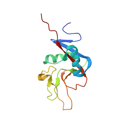
Structure of the Complex of F-Actin and DNGR-1, a C-Type Lectin Receptor Involved in Dendritic Cell Cross-Presentation of Dead Cell-Associated Antigens.

Hanc, P.Fujii, T.Iborra, S.Yamada, Y.Huotari, J.Schulz, O.Ahrens, S.Kjer, S.Way, M.Sancho, D.Namba, K.Reis e Sousa, C.

(2015) Immunity 42: 839-849

  • DOI: https://doi.org/10.1016/j.immuni.2015.04.009
  • Primary Citation of Related Structures:  
    3J82

  • PubMed Abstract: 

    DNGR-1 is a C-type lectin receptor that binds F-actin exposed by dying cells and facilitates cross-presentation of dead cell-associated antigens by dendritic cells. Here we present the structure of DNGR-1 bound to F-actin at 7.7 Å resolution. Unusually for F-actin binding proteins, the DNGR-1 ligand binding domain contacts three actin subunits helically arranged in the actin filament, bridging over two protofilaments, as well as two neighboring actin subunits along one protofilament. Mutation of residues predicted to mediate ligand binding led to loss of DNGR-1-dependent cross-presentation of dead cell-associated antigens, formally demonstrating that the latter depends on F-actin recognition. Notably, DNGR-1 has relatively modest affinity for F-actin but multivalent interactions allow a marked increase in binding strength. Our findings shed light on modes of actin binding by cellular proteins and reveal how extracellular detection of cytoskeletal components by dedicated receptors allows immune monitoring of loss of cellular integrity.


  • Organizational Affiliation

    Immunobiology Laboratory, The Francis Crick Institute, Lincoln's Inn Fields Laboratory, 44 Lincoln's Inn Fields, London WC2A 3LY, UK.


Macromolecules
Find similar proteins by:  (by identity cutoff)  |  3D Structure
Entity ID: 1
MoleculeChains Sequence LengthOrganismDetailsImage
C-type lectin domain family 9 member A131Mus musculusMutation(s): 0 
Gene Names: Clec9aDngr-1
UniProt & NIH Common Fund Data Resources
Find proteins for Q8BRU4 (Mus musculus)
Explore Q8BRU4 
Go to UniProtKB:  Q8BRU4
IMPC:  MGI:2444608
Entity Groups  
Sequence Clusters30% Identity50% Identity70% Identity90% Identity95% Identity100% Identity
UniProt GroupQ8BRU4
Sequence Annotations
Expand
  • Reference Sequence
Find similar proteins by:  (by identity cutoff)  |  3D Structure
Entity ID: 2
MoleculeChains Sequence LengthOrganismDetailsImage
Actin, cytoplasmic 1
B, C, D
374Homo sapiensMutation(s): 0 
UniProt & NIH Common Fund Data Resources
Find proteins for P60709 (Homo sapiens)
Explore P60709 
Go to UniProtKB:  P60709
PHAROS:  P60709
GTEx:  ENSG00000075624 
Entity Groups  
Sequence Clusters30% Identity50% Identity70% Identity90% Identity95% Identity100% Identity
UniProt GroupP60709
Sequence Annotations
Expand
  • Reference Sequence
Experimental Data & Validation

Experimental Data

  • Method: ELECTRON MICROSCOPY
  • Resolution: 7.70 Å
  • Aggregation State: FILAMENT 
  • Reconstruction Method: SINGLE PARTICLE 
EM Software:
TaskSoftware PackageVersion
RECONSTRUCTIONEMAN
RECONSTRUCTIONSPIDER

Structure Validation

View Full Validation Report


Entry History 

Deposition Data

Revision History  (Full details and data files)

  • Version 1.0: 2015-05-20
    Type: Initial release
  • Version 1.1: 2015-05-27
    Changes: Database references, Other
  • Version 1.2: 2019-12-18
    Changes: Author supporting evidence, Data collection, Database references, Derived calculations