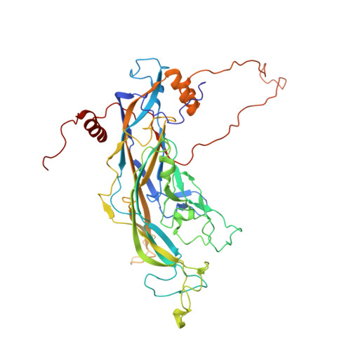
Maturation of the human papillomavirus 16 capsid.
Cardone, G., Moyer, A.L., Cheng, N., Thompson, C.D., Dvoretzky, I., Lowy, D.R., Schiller, J.T., Steven, A.C., Buck, C.B., Trus, B.L.(2014) mBio 5: e01104-e01114
- PubMed: 25096873
- DOI: https://doi.org/10.1128/mBio.01104-14
- Primary Citation of Related Structures:
3J6R - PubMed Abstract:
Papillomaviruses are a family of nonenveloped DNA viruses that infect the skin or mucosa of their vertebrate hosts. The viral life cycle is closely tied to the differentiation of infected keratinocytes. Papillomavirus virions are released into the environment through a process known as desquamation, in which keratinocytes lose structural integrity prior to being shed from the surface of the skin. During this process, virions are exposed to an increasingly oxidative environment, leading to their stabilization through the formation of disulfide cross-links between neighboring molecules of the major capsid protein, L1. We used time-lapse cryo-electron microscopy and image analysis to study the maturation of HPV16 capsids assembled in mammalian cells and exposed to an oxidizing environment after cell lysis. Initially, the virion is a loosely connected procapsid that, under in vitro conditions, condenses over several hours into the more familiar 60-nm-diameter papillomavirus capsid. In this process, the procapsid shrinks by ~5% in diameter, its pentameric capsomers change in structure (most markedly in the axial region), and the interaction surfaces between adjacent capsomers are consolidated. A C175S mutant that cannot achieve normal inter-L1 disulfide cross-links shows maturation-related shrinkage but does not achieve the fully condensed 60-nm form. Pseudoatomic modeling based on a 9-Å resolution reconstruction of fully mature capsids revealed C-terminal disulfide-stabilized "suspended bridges" that form intercapsomeric cross-links. The data suggest a model in which procapsids exist in a range of dynamic intermediates that can be locked into increasingly mature configurations by disulfide cross-linking, possibly through a Brownian ratchet mechanism. Importance: Human papillomaviruses (HPVs) cause nearly all cases of cervical cancer, a major fraction of cancers of the penis, vagina/vulva, anus, and tonsils, and genital and nongenital warts. HPV types associated with a high risk of cancer, such as HPV16, are generally transmitted via sexual contact. The nonenveloped virion of HPVs shows a high degree of stability, allowing the virus to persist in an infectious form in environmental fomites. In this study, we used cryo-electron microscopy to elucidate the structure of the HPV16 capsid at different stages of maturation. The fully mature capsid adopts a rigid, highly regular structure stabilized by intermolecular disulfide bonds. The availability of a pseudoatomic model of the fully mature HPV16 virion should help guide understanding of antibody responses elicited by HPV capsid-based vaccines.
Organizational Affiliation:
Laboratory of Structural Biology, National Institute for Arthritis, Musculoskeletal and Skin Diseases, National Institutes of Health (NIH), Bethesda, Maryland, USA.