3ITJ

Crystal structure of Saccharomyces cerevisiae thioredoxin reductase 1 (Trr1)


Experimental Data Snapshot

  • Method: X-RAY DIFFRACTION
  • Resolution: 2.40 Å
  • R-Value Free: 0.194 
  • R-Value Work: 0.169 
  • R-Value Observed: 0.171 

wwPDB Validation   3D Report Full Report


Ligand Structure Quality Assessment 


This is version 1.2 of the entry. See complete history


Literature

Insights into the specificity of thioredoxin reductase-thioredoxin interactions. A structural and functional investigation of the yeast thioredoxin system.

Oliveira, M.A.Discola, K.F.Alves, S.V.Medrano, F.J.Guimaraes, B.G.Netto, L.E.

(2010) Biochemistry 49: 3317-3326

  • DOI: https://doi.org/10.1021/bi901962p
  • Primary Citation of Related Structures:  
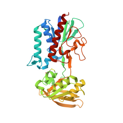
    3ITJ

  • PubMed Abstract: 

    The enzymatic activity of thioredoxin reductase enzymes is endowed by at least two redox centers: a flavin and a dithiol/disulfide CXXC motif. The interaction between thioredoxin reductase and thioredoxin is generally species-specific, but the molecular aspects related to this phenomenon remain elusive. Here, we investigated the yeast cytosolic thioredoxin system, which is composed of NADPH, thioredoxin reductase (ScTrxR1), and thioredoxin 1 (ScTrx1) or thioredoxin 2 (ScTrx2). We showed that ScTrxR1 was able to efficiently reduce yeast thioredoxins (mitochondrial and cytosolic) but failed to reduce the human and Escherichia coli thioredoxin counterparts. To gain insights into this specificity, the crystallographic structure of oxidized ScTrxR1 was solved at 2.4 A resolution. The protein topology of the redox centers indicated the necessity of a large structural rearrangement for FAD and thioredoxin reduction using NADPH. Therefore, we modeled a large structural rotation between the two ScTrxR1 domains (based on the previously described crystal structure, PDB code 1F6M ). Employing diverse approaches including enzymatic assays, site-directed mutagenesis, amino acid sequence alignment, and structure comparisons, insights were obtained about the features involved in the species-specificity phenomenon, such as complementary electronic parameters between the surfaces of ScTrxR1 and yeast thioredoxin enzymes and loops and residues (such as Ser(72) in ScTrx2). Finally, structural comparisons and amino acid alignments led us to propose a new classification that includes a larger number of enzymes with thioredoxin reductase activity, neglected in the low/high molecular weight classification.


  • Organizational Affiliation

    Departamento de Biologia, Universidade Estadual Paulista, São Vicente, Brazil.


Macromolecules
Find similar proteins by:  (by identity cutoff)  |  3D Structure
Entity ID: 1
MoleculeChains Sequence LengthOrganismDetailsImage
Thioredoxin reductase 1
A, B, C, D
338Saccharomyces cerevisiae W303Mutation(s): 0 
Gene Names: D9476.5TRR1YDR353W
EC: 1.8.1.9
UniProt
Find proteins for P29509 (Saccharomyces cerevisiae (strain ATCC 204508 / S288c))
Explore P29509 
Go to UniProtKB:  P29509
Entity Groups  
Sequence Clusters30% Identity50% Identity70% Identity90% Identity95% Identity100% Identity
UniProt GroupP29509
Sequence Annotations
Expand
  • Reference Sequence
Small Molecules
Ligands 2 Unique
IDChains Name / Formula / InChI Key2D Diagram3D Interactions
FAD
Query on FAD

Download Ideal Coordinates CCD File 
E [auth A],
G [auth B],
J [auth C],
M [auth D]
FLAVIN-ADENINE DINUCLEOTIDE
C27 H33 N9 O15 P2
VWWQXMAJTJZDQX-UYBVJOGSSA-N
CIT
Query on CIT

Download Ideal Coordinates CCD File 
F [auth A]
H [auth B]
I [auth B]
K [auth C]
L [auth C]
F [auth A],
H [auth B],
I [auth B],
K [auth C],
L [auth C],
N [auth D]
CITRIC ACID
C6 H8 O7
KRKNYBCHXYNGOX-UHFFFAOYSA-N
Experimental Data & Validation

Experimental Data

  • Method: X-RAY DIFFRACTION
  • Resolution: 2.40 Å
  • R-Value Free: 0.194 
  • R-Value Work: 0.169 
  • R-Value Observed: 0.171 
  • Space Group: C 1 2 1
Unit Cell:
Length ( Å )Angle ( ˚ )
a = 127.975α = 90
b = 135.411β = 89.95
c = 75.819γ = 90
Software Package:
Software NamePurpose
MAR345dtbdata collection
AMoREphasing
REFMACrefinement
MOSFLMdata reduction
SCALAdata scaling

Structure Validation

View Full Validation Report


Ligand Structure Quality Assessment 


Entry History 

Deposition Data

Revision History  (Full details and data files)

  • Version 1.0: 2010-03-31
    Type: Initial release
  • Version 1.1: 2011-07-13
    Changes: Advisory, Refinement description, Source and taxonomy, Version format compliance
  • Version 1.2: 2023-09-06
    Changes: Data collection, Database references, Derived calculations, Refinement description