


Crystal structure of Helicobacter pylori thioredoxin reductase
Sanders, D., Obiero, J., van Straaten, K.To be published.
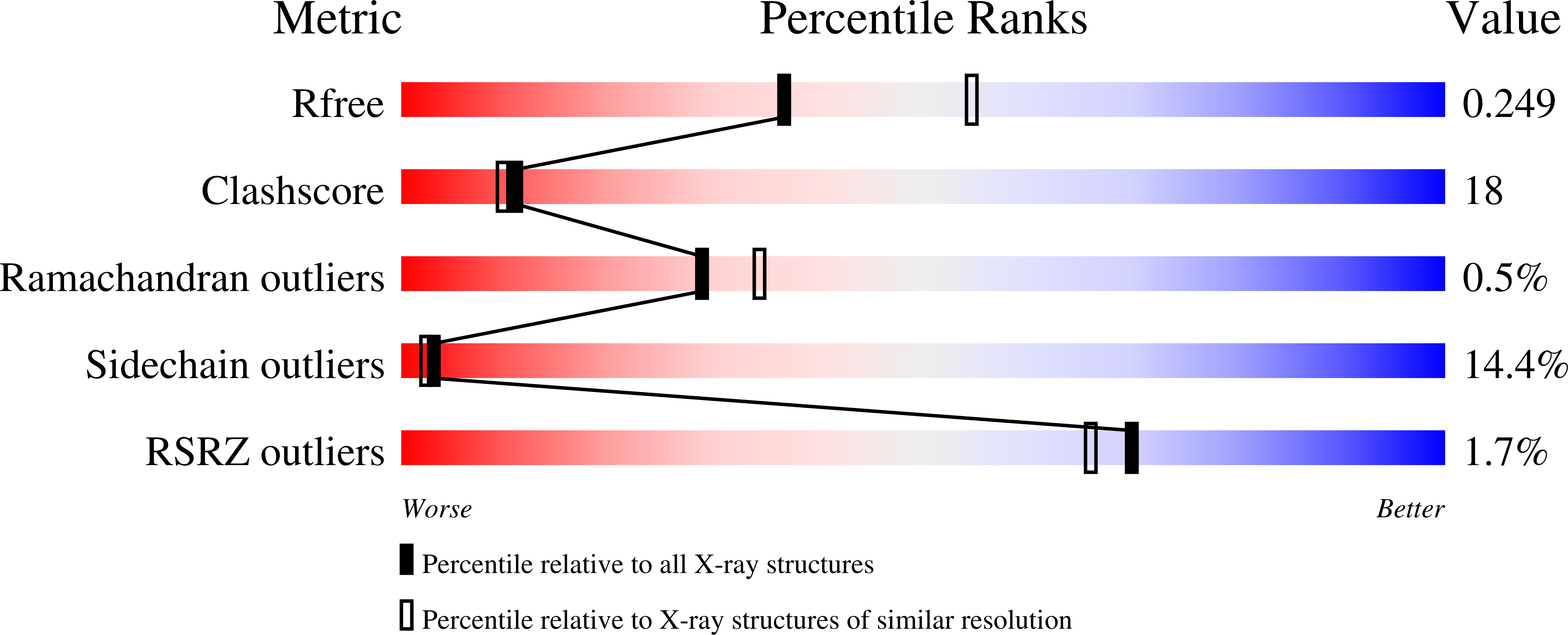
Experimental Data Snapshot
Entity ID: 1 | |||||
|---|---|---|---|---|---|
| Molecule | Chains | Sequence Length | Organism | Details | Image |
| Thioredoxin reductase | 311 | Helicobacter pylori 26695 | Mutation(s): 0 Gene Names: HP0825 | 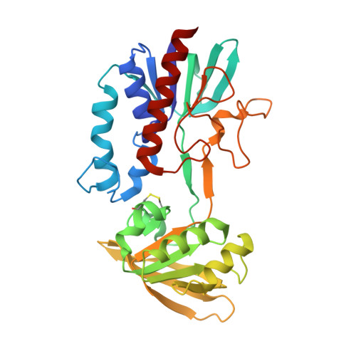 | |
UniProt | |||||
Find proteins for P56431 (Helicobacter pylori (strain ATCC 700392 / 26695)) Explore P56431 Go to UniProtKB: P56431 | |||||
Entity Groups | |||||
| Sequence Clusters | 30% Identity50% Identity70% Identity90% Identity95% Identity100% Identity | ||||
| UniProt Group | P56431 | ||||
Sequence AnnotationsExpand | |||||
| |||||
| Ligands 1 Unique | |||||
|---|---|---|---|---|---|
| ID | Chains | Name / Formula / InChI Key | 2D Diagram | 3D Interactions | |
| FAD Query on FAD | D [auth A], E [auth B], F [auth C] | FLAVIN-ADENINE DINUCLEOTIDE C27 H33 N9 O15 P2 VWWQXMAJTJZDQX-UYBVJOGSSA-N |  | ||
| Length ( Å ) | Angle ( ˚ ) |
|---|---|
| a = 89.4 | α = 90 |
| b = 89.4 | β = 90 |
| c = 279.9 | γ = 120 |
| Software Name | Purpose |
|---|---|
| XDS | data scaling |
| MrBUMP | phasing |
| REFMAC | refinement |
| XDS | data reduction |
RCSB PDB (citation) is hosted by
RCSB PDB is a member of the

