3IS2

2.3 Angstrom Crystal Structure of a Cys71 Sulfenic Acid form of Vivid


Experimental Data Snapshot

  • Method: X-RAY DIFFRACTION
  • Resolution: 2.30 Å
  • R-Value Free: 0.254 
  • R-Value Work: 0.226 
  • R-Value Observed: 0.254 

wwPDB Validation   3D Report Full Report


Ligand Structure Quality Assessment 


This is version 1.1 of the entry. See complete history


Literature

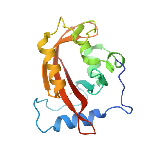
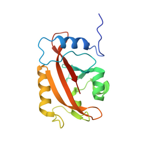
Illuminating solution responses of a LOV domain protein with photocoupled small-angle X-ray scattering.

Lamb, J.S.Zoltowski, B.D.Pabit, S.A.Li, L.Crane, B.R.Pollack, L.

(2009) J Mol Biol 393: 909-919

  • DOI: https://doi.org/10.1016/j.jmb.2009.08.045
  • Primary Citation of Related Structures:  
    3IS2

  • PubMed Abstract: 

    The PAS-LOV domain is a signal-transducing component found in a large variety of proteins that is responsible for sensing different stimuli such as light, oxygen, and voltage. The LOV protein VVD regulates blue light responses in the filamentous fungi Neurospora crassa. Using photocoupled, time-resolved small-angle X-ray scattering, we extract the solution protein structure in both dark-adapted and light-activated states. Two distinct dark-adapted conformations are detected in the wild-type protein: a compact structure that corresponds to the crystal structure of the dark-state monomer as well as an extended structure that is well modeled by introducing conformational disorder at the N-terminus of the protein. These conformations are accentuated in carefully selected variants, in which a key residue for propagating structural transitions, Cys71, has been mutated or oxidized. Despite different dark-state conformations, all proteins form a common dimer in response to illumination. Taken together, these data support a reaction scheme that describes the mechanism for light-induced dimerization of VVD. Envelope reconstructions of the transient light-state dimer reveal structures that are best described by a parallel arrangement of subunits that have significantly changed conformation compared to the crystal structure.


  • Organizational Affiliation

    School of Applied and Engineering Physics, Cornell University, Ithaca, NY 14853, USA.


Macromolecules
Find similar proteins by:  (by identity cutoff)  |  3D Structure
Entity ID: 1
MoleculeChains Sequence LengthOrganismDetailsImage
Vivid PAS protein VVD154Neurospora crassaMutation(s): 0 
Gene Names: G17A4.050Vividvvd
UniProt
Find proteins for Q9C3Y6 (Neurospora crassa)
Explore Q9C3Y6 
Go to UniProtKB:  Q9C3Y6
Entity Groups  
Sequence Clusters30% Identity50% Identity70% Identity90% Identity95% Identity100% Identity
UniProt GroupQ9C3Y6
Sequence Annotations
Expand
  • Reference Sequence
Find similar proteins by:  (by identity cutoff)  |  3D Structure
Entity ID: 2
MoleculeChains Sequence LengthOrganismDetailsImage
Vivid PAS protein VVD154Neurospora crassaMutation(s): 0 
Gene Names: G17A4.050Vividvvd
UniProt
Find proteins for Q9C3Y6 (Neurospora crassa)
Explore Q9C3Y6 
Go to UniProtKB:  Q9C3Y6
Entity Groups  
Sequence Clusters30% Identity50% Identity70% Identity90% Identity95% Identity100% Identity
UniProt GroupQ9C3Y6
Sequence Annotations
Expand
  • Reference Sequence
Small Molecules
Ligands 1 Unique
IDChains Name / Formula / InChI Key2D Diagram3D Interactions
FAD
Query on FAD

Download Ideal Coordinates CCD File 
C [auth A],
D [auth B]
FLAVIN-ADENINE DINUCLEOTIDE
C27 H33 N9 O15 P2
VWWQXMAJTJZDQX-UYBVJOGSSA-N
Modified Residues  1 Unique
IDChains TypeFormula2D DiagramParent
CSO
Query on CSO
A
L-PEPTIDE LINKINGC3 H7 N O3 SCYS
Experimental Data & Validation

Experimental Data

  • Method: X-RAY DIFFRACTION
  • Resolution: 2.30 Å
  • R-Value Free: 0.254 
  • R-Value Work: 0.226 
  • R-Value Observed: 0.254 
  • Space Group: P 1 21 1
Unit Cell:
Length ( Å )Angle ( ˚ )
a = 36.42α = 90
b = 81.11β = 95.03
c = 58.03γ = 90
Software Package:
Software NamePurpose
HKL-2000data collection
AMoREphasing
CNSrefinement
HKL-2000data reduction
HKL-2000data scaling

Structure Validation

View Full Validation Report


Ligand Structure Quality Assessment 


Entry History 

Deposition Data

Revision History  (Full details and data files)

  • Version 1.0: 2009-11-03
    Type: Initial release
  • Version 1.1: 2011-07-13
    Changes: Version format compliance