3IMP

New crystal form of the C-terminal domain of Helicobacter pylori MotB (residues 125-256)


Experimental Data Snapshot

  • Method: X-RAY DIFFRACTION
  • Resolution: 2.50 Å
  • R-Value Free: 0.249 
  • R-Value Work: 0.186 
  • R-Value Observed: 0.189 

wwPDB Validation   3D Report Full Report


This is version 1.2 of the entry. See complete history


Literature

Crystallographic and Molecular Dynamics Analysis of Loop Motions Unmasking the Peptidoglycan-Binding Site in Stator Protein MotB of Flagellar Motor

Reboul, C.F.Andrews, D.A.Nahar, M.F.Buckle, A.M.Roujeinikova, A.

(2011) PLoS One 6: e18981-e18981

  • DOI: https://doi.org/10.1371/journal.pone.0018981
  • Primary Citation of Related Structures:  
    3IMP

  • PubMed Abstract: 

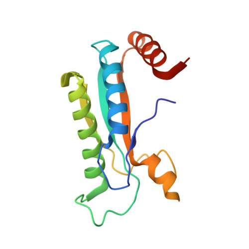
    The C-terminal domain of MotB (MotB-C) shows high sequence similarity to outer membrane protein A and related peptidoglycan (PG)-binding proteins. It is believed to anchor the power-generating MotA/MotB stator unit of the bacterial flagellar motor to the peptidoglycan layer of the cell wall. We previously reported the first crystal structure of this domain and made a puzzling observation that all conserved residues that are thought to be essential for PG recognition are buried and inaccessible in the crystal structure. In this study, we tested a hypothesis that peptidoglycan binding is preceded by, or accompanied by, some structural reorganization that exposes the key conserved residues.


  • Organizational Affiliation

    Department of Microbiology and Department of Biochemistry and Molecular Biology, Monash University, Clayton, Victoria, Australia.


Macromolecules
Find similar proteins by:  (by identity cutoff)  |  3D Structure
Entity ID: 1
MoleculeChains Sequence LengthOrganismDetailsImage
Chemotaxis protein motB138Helicobacter pylori 26695Mutation(s): 0 
Gene Names: motB
UniProt
Find proteins for P56427 (Helicobacter pylori (strain ATCC 700392 / 26695))
Explore P56427 
Go to UniProtKB:  P56427
Entity Groups  
Sequence Clusters30% Identity50% Identity70% Identity90% Identity95% Identity100% Identity
UniProt GroupP56427
Sequence Annotations
Expand
  • Reference Sequence
Experimental Data & Validation

Experimental Data

  • Method: X-RAY DIFFRACTION
  • Resolution: 2.50 Å
  • R-Value Free: 0.249 
  • R-Value Work: 0.186 
  • R-Value Observed: 0.189 
  • Space Group: P 1 21 1
Unit Cell:
Length ( Å )Angle ( ˚ )
a = 107.56α = 90
b = 100.336β = 119.51
c = 108.49γ = 90
Software Package:
Software NamePurpose
REFMACrefinement
PDB_EXTRACTdata extraction
MOSFLMdata reduction
SCALAdata scaling
AMoREphasing

Structure Validation

View Full Validation Report


Entry History 

Deposition Data

Revision History  (Full details and data files)

  • Version 1.0: 2010-08-04
    Type: Initial release
  • Version 1.1: 2011-07-13
    Changes: Version format compliance
  • Version 1.2: 2023-11-01
    Changes: Data collection, Database references, Derived calculations, Refinement description