3IF6

Crystal structure of OXA-46 beta-lactamase from P. aeruginosa


Experimental Data Snapshot

  • Method: X-RAY DIFFRACTION
  • Resolution: 2.40 Å
  • R-Value Free: 0.285 
  • R-Value Work: 0.211 
  • R-Value Observed: 0.218 

wwPDB Validation   3D Report Full Report


This is version 1.4 of the entry. See complete history


Literature

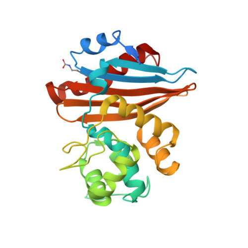
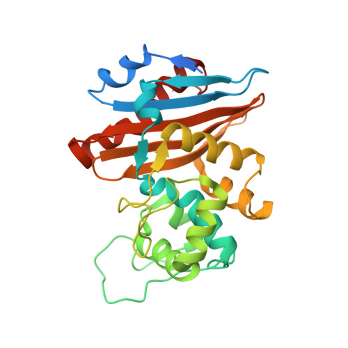
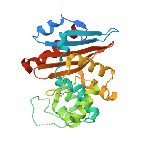
Crystal structure of the narrow-spectrum OXA-46 class D beta-lactamase: relationship between active-site lysine carbamylation and inhibition by polycarboxylates

Docquier, J.D.Benvenuti, M.Calderone, V.Giuliani, F.Kapetis, D.De Luca, F.Rossolini, G.M.Mangani, S.

(2010) Antimicrob Agents Chemother 54: 2167-2174

  • DOI: https://doi.org/10.1128/AAC.01517-09
  • Primary Citation of Related Structures:  
    3IF6

  • PubMed Abstract: 

    Class D beta-lactamases represent a heterogeneous group of active-site serine beta-lactamases that show an extraordinary panel of functional features and substrate profiles, thus representing relevant models for biochemical and structural studies. OXA-46 is a narrow-spectrum enzyme belonging to the OXA-2 subgroup which was found in a Pseudomonas aeruginosa clinical isolate from northern Italy. In this work, we obtained the three-dimensional structure of OXA-46, which shows the overall fold of active serine beta-lactamases and a dimeric quaternary structure. Significant differences with currently available structures of class D beta-lactamases were found in the loops located close to the active site, which differ in length and conformation. Interestingly, the three subunits present in the asymmetric unit showed some structural heterogeneity, only one of which presented a carbamylated lysine recognized as an important functional feature of class D enzymes. The carbamylation state of residue Lys75 appeared to be associated with different shapes and dimensions of the active site. Moreover, a tartrate molecule from the crystallization buffer was found in the active site of the noncarbamylated subunits, which interacts with catalytically relevant residues. The OXA-46 crystal asymmetric units thus interestingly present the structures of the free carbamylated active site and of the ligand-bound uncarbamylated active site, offering the structural basis for investigating the potential of new scaffolds of beta-lactamase inhibitors.


  • Organizational Affiliation

    Dipartimento di Biologia Molecolare, Università di Siena, Siena, Italy.


Macromolecules
Find similar proteins by:  (by identity cutoff)  |  3D Structure
Entity ID: 1
MoleculeChains Sequence LengthOrganismDetailsImage
OXA-46 oxacillinase266Pseudomonas aeruginosaMutation(s): 0 
UniProt
Find proteins for Q8GRH0 (Pseudomonas aeruginosa)
Explore Q8GRH0 
Go to UniProtKB:  Q8GRH0
Entity Groups  
Sequence Clusters30% Identity50% Identity70% Identity90% Identity95% Identity100% Identity
UniProt GroupQ8GRH0
Sequence Annotations
Expand
  • Reference Sequence
Find similar proteins by:  (by identity cutoff)  |  3D Structure
Entity ID: 2
MoleculeChains Sequence LengthOrganismDetailsImage
OXA-46 oxacillinase266Pseudomonas aeruginosaMutation(s): 0 
UniProt
Find proteins for Q8GRH0 (Pseudomonas aeruginosa)
Explore Q8GRH0 
Go to UniProtKB:  Q8GRH0
Entity Groups  
Sequence Clusters30% Identity50% Identity70% Identity90% Identity95% Identity100% Identity
UniProt GroupQ8GRH0
Sequence Annotations
Expand
  • Reference Sequence
Find similar proteins by:  (by identity cutoff)  |  3D Structure
Entity ID: 3
MoleculeChains Sequence LengthOrganismDetailsImage
OXA-46 oxacillinase266Pseudomonas aeruginosaMutation(s): 0 
UniProt
Find proteins for Q8GRH0 (Pseudomonas aeruginosa)
Explore Q8GRH0 
Go to UniProtKB:  Q8GRH0
Entity Groups  
Sequence Clusters30% Identity50% Identity70% Identity90% Identity95% Identity100% Identity
UniProt GroupQ8GRH0
Sequence Annotations
Expand
  • Reference Sequence
Small Molecules
Ligands 3 Unique
IDChains Name / Formula / InChI Key2D Diagram3D Interactions
P6G
Query on P6G

Download Ideal Coordinates CCD File 
H [auth B]HEXAETHYLENE GLYCOL
C12 H26 O7
IIRDTKBZINWQAW-UHFFFAOYSA-N
TLA
Query on TLA

Download Ideal Coordinates CCD File 
D [auth A],
G [auth B]
L(+)-TARTARIC ACID
C4 H6 O6
FEWJPZIEWOKRBE-JCYAYHJZSA-N
EDO
Query on EDO

Download Ideal Coordinates CCD File 
E [auth A]
F [auth A]
I [auth B]
J [auth B]
K [auth B]
E [auth A],
F [auth A],
I [auth B],
J [auth B],
K [auth B],
L [auth B]
1,2-ETHANEDIOL
C2 H6 O2
LYCAIKOWRPUZTN-UHFFFAOYSA-N
Modified Residues  1 Unique
IDChains TypeFormula2D DiagramParent
KCX
Query on KCX
A
L-PEPTIDE LINKINGC7 H14 N2 O4LYS
Experimental Data & Validation

Experimental Data

  • Method: X-RAY DIFFRACTION
  • Resolution: 2.40 Å
  • R-Value Free: 0.285 
  • R-Value Work: 0.211 
  • R-Value Observed: 0.218 
  • Space Group: H 3 2
Unit Cell:
Length ( Å )Angle ( ˚ )
a = 123.838α = 90
b = 123.838β = 90
c = 327.915γ = 120
Software Package:
Software NamePurpose
MOLREPphasing
REFMACrefinement
MOSFLMdata reduction
SCALAdata scaling

Structure Validation

View Full Validation Report


Entry History 

Deposition Data

Revision History  (Full details and data files)

  • Version 1.0: 2010-03-16
    Type: Initial release
  • Version 1.1: 2011-07-13
    Changes: Version format compliance
  • Version 1.2: 2014-02-19
    Changes: Database references
  • Version 1.3: 2023-11-01
    Changes: Data collection, Database references, Derived calculations, Refinement description
  • Version 1.4: 2023-11-22
    Changes: Data collection