3I27

Structure of bovine torovirus Hemagglutinin-Esterase in complex with receptor


Experimental Data Snapshot

  • Method: X-RAY DIFFRACTION
  • Resolution: 2.00 Å
  • R-Value Free: 0.210 
  • R-Value Work: 0.179 
  • R-Value Observed: 0.180 

wwPDB Validation   3D Report Full Report


Ligand Structure Quality Assessment 


This is version 2.1 of the entry. See complete history


Literature

Structural basis for ligand and substrate recognition by torovirus hemagglutinin esterases

Langereis, M.A.Zeng, Q.H.Gerwig, G.J.Frey, B.von Itzstein, M.Kamerling, J.P.de Groot, R.J.Huizinga, E.G.

(2009) Proc Natl Acad Sci U S A 106: 15897-15902

  • DOI: https://doi.org/10.1073/pnas.0904266106
  • Primary Citation of Related Structures:  
    3I1K, 3I1L, 3I26, 3I27

  • PubMed Abstract: 

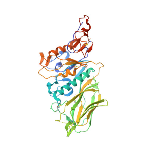
    Hemagglutinin esterases (HEs), closely related envelope glycoproteins in influenza C and corona- and toroviruses, mediate reversible attachment to O-acetylated sialic acids (Sias). They do so by acting both as lectins and as receptor-destroying enzymes, functions exerted by separate protein domains. HE divergence was accompanied by changes in quaternary structure and in receptor and substrate specificity. The selective forces underlying HE diversity and the molecular basis for Sia specificity are poorly understood. Here we present crystal structures of porcine and bovine torovirus HEs in complex with receptor analogs. Torovirus HEs form homodimers with sialate-O-acetylesterase domains almost identical to corresponding domains in orthomyxo- and coronavirus HEs, but with unique lectin sites. Structure-guided biochemical analysis of the esterase domains revealed that a functionally, but not structurally conserved arginine-Sia carboxylate interaction is critical for the binding and positioning of glycosidically bound Sias in the catalytic pocket. Although essential for efficient de-O-acetylation of Sias, this interaction is not required for catalysis nor does it affect substrate specificity. In fact, the distinct preference of the porcine torovirus enzyme for 9-mono- over 7,9-di-O-acetylated Sias can be explained from a single-residue difference with HEs of more promiscuous specificity. Apparently, esterase and lectin pockets coevolved; also the porcine torovirus HE receptor-binding site seems to have been designed to use 9-mono- and exclude di-O-acetylated Sias, possibly as an adaptation to replication in swine. Our findings shed light on HE evolution and provide fundamental insight into mechanisms of substrate binding, substrate recognition, and receptor selection in this important class of virion proteins.


  • Organizational Affiliation

    Virology Division, Department of Infectious Diseases & Immunology, Faculty of Veterinary Medicine, Utrecht University, 3584 CH Utrecht, The Netherlands.


Macromolecules
Find similar proteins by:  (by identity cutoff)  |  3D Structure
Entity ID: 1
MoleculeChains Sequence LengthOrganismDetailsImage
Hemagglutinin-esterase
A, B, C, D
384Breda virus serotype 1Mutation(s): 0 
Gene Names: HE
EC: 3.1.1.53
UniProt
Find proteins for P0C0V9 (Breda virus 1)
Explore P0C0V9 
Go to UniProtKB:  P0C0V9
Entity Groups  
Sequence Clusters30% Identity50% Identity70% Identity90% Identity95% Identity100% Identity
UniProt GroupP0C0V9
Sequence Annotations
Expand
  • Reference Sequence
Oligosaccharides

Help

Entity ID: 2
MoleculeChains Length2D Diagram Glycosylation3D Interactions
beta-D-mannopyranose-(1-4)-2-acetamido-2-deoxy-beta-D-glucopyranose-(1-4)-2-acetamido-2-deoxy-beta-D-glucopyranose
E, H, J
3N-Glycosylation
Glycosylation Resources
GlyTouCan:  G15407YE
GlyCosmos:  G15407YE
GlyGen:  G15407YE
Entity ID: 3
MoleculeChains Length2D Diagram Glycosylation3D Interactions
2-acetamido-2-deoxy-beta-D-glucopyranose-(1-4)-2-acetamido-2-deoxy-beta-D-glucopyranose
F, G, I, K, L
2N-Glycosylation
Glycosylation Resources
GlyTouCan:  G42666HT
GlyCosmos:  G42666HT
GlyGen:  G42666HT
Entity ID: 4
MoleculeChains Length2D Diagram Glycosylation3D Interactions
alpha-D-mannopyranose-(1-3)-[alpha-D-mannopyranose-(1-6)]alpha-D-mannopyranose-(1-6)-[alpha-D-mannopyranose-(1-3)]beta-D-mannopyranose-(1-4)-2-acetamido-2-deoxy-beta-D-glucopyranose-(1-4)-2-acetamido-2-deoxy-beta-D-glucopyranose
M
7N-Glycosylation
Glycosylation Resources
GlyTouCan:  G55220VL
GlyCosmos:  G55220VL
GlyGen:  G55220VL
Experimental Data & Validation

Experimental Data

  • Method: X-RAY DIFFRACTION
  • Resolution: 2.00 Å
  • R-Value Free: 0.210 
  • R-Value Work: 0.179 
  • R-Value Observed: 0.180 
  • Space Group: P 21 21 21
Unit Cell:
Length ( Å )Angle ( ˚ )
a = 67.63α = 90
b = 112.89β = 90
c = 273.03γ = 90
Software Package:
Software NamePurpose
DNAdata collection
PHASERphasing
REFMACrefinement
XDSdata reduction
SCALAdata scaling

Structure Validation

View Full Validation Report


Ligand Structure Quality Assessment 


Entry History 

Deposition Data

Revision History  (Full details and data files)

  • Version 1.0: 2009-09-15
    Type: Initial release
  • Version 1.1: 2011-07-13
    Changes: Advisory, Version format compliance
  • Version 2.0: 2020-07-29
    Type: Remediation
    Reason: Carbohydrate remediation
    Changes: Advisory, Atomic model, Data collection, Database references, Derived calculations, Structure summary
  • Version 2.1: 2023-11-01
    Changes: Data collection, Database references, Refinement description, Structure summary