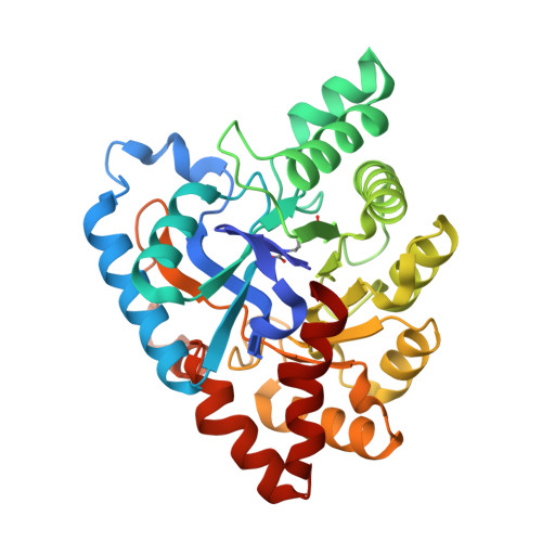
Structure-based and random mutagenesis approaches increase the organophosphate-degrading activity of a phosphotriesterase homologue from Deinococcus radiodurans.
Hawwa, R., Larsen, S.D., Ratia, K., Mesecar, A.D.(2009) J Mol Biol 393: 36-57
- PubMed: 19631223
- DOI: https://doi.org/10.1016/j.jmb.2009.06.083
- Primary Citation of Related Structures:
3GTF, 3GTH, 3GTI, 3GTX, 3GU1, 3GU2, 3GU9, 3HTW - PubMed Abstract:
An enzyme from the amidohydrolase family from Deinococcus radiodurans (Dr-OPH) with homology to phosphotriesterase has been shown to exhibit activity against both organophosphate (OP) and lactone compounds. We have characterized the physical properties of Dr-OPH and have found it to be a highly thermostable enzyme, remaining active after 3 h of incubation at 60 degrees C and withstanding incubation at temperatures up to 70 degrees C. In addition, it can withstand concentrations of at least 200 mg/mL. These properties make Dr-OPH a promising candidate for development in commercial applications. However, compared to the most widely studied OP-degrading enzyme, that from Pseudomonas diminuta, Dr-OPH has low hydrolytic activity against certain OP substrates. Therefore, we sought to improve the OP-degrading activity of Dr-OPH, specifically toward the pesticides ethyl and methyl paraoxon, using structure-based and random approaches. Site-directed mutagenesis, random mutagenesis, and site-saturation mutagenesis were utilized to increase the OP-degrading activity of Dr-OPH. Out of a screen of more than 30,000 potential mutants, a total of 26 mutant enzymes were purified and characterized kinetically. Crystal structures of w.t. Dr-OPH, of Dr-OPH in complex with a product analog, and of 7 mutant enzymes were determined to resolutions between 1.7 and 2.4 A. Information from these structures directed the design and production of 4 additional mutants for analysis. In total, our mutagenesis efforts improved the catalytic activity of Dr-OPH toward ethyl and methyl paraoxon by 126- and 322-fold and raised the specificity for these two substrates by 557- and 183-fold, respectively. Our work highlights the importance of an iterative approach to mutagenesis, proving that large rate enhancements are achieved when mutations are made in already active mutants. In addition, the relationship between the kinetic parameters and the introduced mutations has allowed us to hypothesize on those factors most important for maintaining the structure and function of the enzyme.
Organizational Affiliation:
Department of Medicinal Chemistry and Pharmacognosy, Center for Pharmaceutical Biotechnology, University of Illinois at Chicago, 900 South Ashland Avenue, Chicago, IL 60607, USA.