


Crystal structure of the ligand binding domain of E. coli CynR with its specific effector azide
Singer, A.U., Evdokimova, E., Kagan, O., Cuff, M.E., Edwards, A.M., Joachimiak, A., Savchenko, A.To be published.
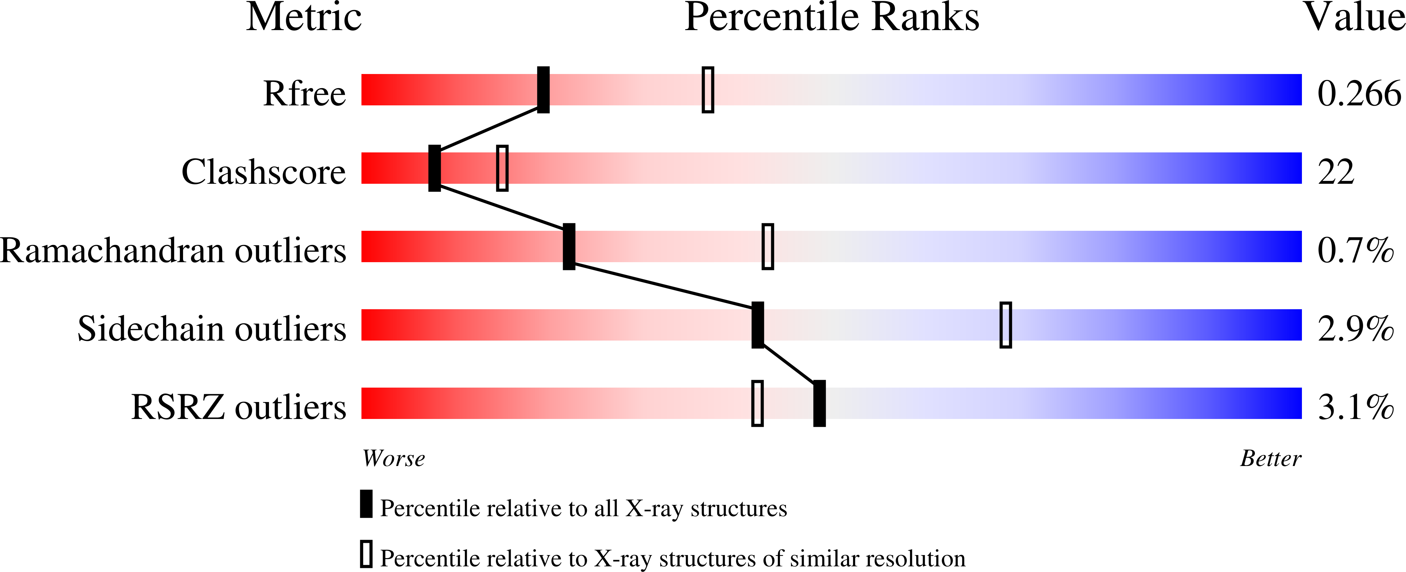
Experimental Data Snapshot
wwPDB Validation 3D Report Full Report
Entity ID: 1 | |||||
|---|---|---|---|---|---|
| Molecule | Chains | Sequence Length | Organism | Details | Image |
| HTH-type transcriptional regulator cynR | 238 | Escherichia coli K-12 | Mutation(s): 0 Gene Names: b0338, cynR, JW5894 | 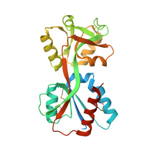 | |
UniProt | |||||
Find proteins for P27111 (Escherichia coli (strain K12)) Explore P27111 Go to UniProtKB: P27111 | |||||
Entity Groups | |||||
| Sequence Clusters | 30% Identity50% Identity70% Identity90% Identity95% Identity100% Identity | ||||
| UniProt Group | P27111 | ||||
Sequence AnnotationsExpand | |||||
| |||||
| Ligands 2 Unique | |||||
|---|---|---|---|---|---|
| ID | Chains | Name / Formula / InChI Key | 2D Diagram | 3D Interactions | |
| EDO Query on EDO | H [auth C], J [auth D] | 1,2-ETHANEDIOL C2 H6 O2 LYCAIKOWRPUZTN-UHFFFAOYSA-N |  | ||
| AZI Query on AZI | E [auth A], F [auth B], G [auth C], I [auth D] | AZIDE ION N3 IVRMZWNICZWHMI-UHFFFAOYSA-N |  | ||
| Modified Residues 1 Unique | |||||
|---|---|---|---|---|---|
| ID | Chains | Type | Formula | 2D Diagram | Parent |
| MSE Query on MSE | A, B, C, D | L-PEPTIDE LINKING | C5 H11 N O2 Se |  | MET |
| Length ( Å ) | Angle ( ˚ ) |
|---|---|
| a = 62.176 | α = 90 |
| b = 98.363 | β = 103.769 |
| c = 86.272 | γ = 90 |
| Software Name | Purpose |
|---|---|
| CrystalClear | data collection |
| AMoRE | phasing |
| CNS | refinement |
| HKL-2000 | data reduction |
| HKL-2000 | data scaling |
RCSB PDB (citation) is hosted by
RCSB PDB is a member of the

