3H3D

Drosophila Pumilio RNA binding domain (Puf domain)


Experimental Data Snapshot

  • Method: X-RAY DIFFRACTION
  • Resolution: 2.30 Å
  • R-Value Free: 0.298 
  • R-Value Work: 0.252 
  • R-Value Observed: 0.254 

wwPDB Validation   3D Report Full Report


This is version 1.3 of the entry. See complete history


Literature

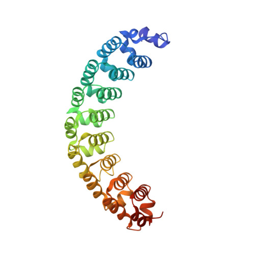
Structure of Pumilio reveals similarity between RNA and peptide binding motifs.

Edwards, T.A.Pyle, S.E.Wharton, R.P.Aggarwal, A.K.

(2001) Cell 105: 281-289

  • DOI: https://doi.org/10.1016/s0092-8674(01)00318-x
  • Primary Citation of Related Structures:  
    3H3D

  • PubMed Abstract: 

    Translation regulation plays an essential role in the differentiation and development of animal cells. One well-studied case is the control of hunchback mRNA during early Drosophila embryogenesis by the trans-acting factors Pumilio, Nanos, and Brain Tumor. We report here a crystal structure of the critical region of Pumilio, the Puf domain, that organizes a multivalent repression complex on the 3' untranslated region of hunchback mRNA. The structure reveals an extended, rainbow shaped molecule, with tandem helical repeats that bear unexpected resemblance to the armadillo repeats in beta-catenin and the HEAT repeats in protein phosphatase 2A. Based on the structure and genetic experiments, we identify putative interaction surfaces for hunchback mRNA and the cofactors Nanos and Brain Tumor. This analysis suggests that similar features in helical repeat proteins are used to bind extended peptides and RNA.


  • Organizational Affiliation

    Structural Biology Program, Department of Physiology and Biophysics, Mount Sinai School of Medicine, Box 1677, 1425 Madison Avenue, New York, NY 10029, USA.


Macromolecules
Find similar proteins by:  (by identity cutoff)  |  3D Structure
Entity ID: 1
MoleculeChains Sequence LengthOrganismDetailsImage
Maternal protein pumilioA [auth X],
B [auth Y]
323Drosophila melanogasterMutation(s): 0 
Gene Names: pumilio
UniProt
Find proteins for P25822 (Drosophila melanogaster)
Explore P25822 
Go to UniProtKB:  P25822
Entity Groups  
Sequence Clusters30% Identity50% Identity70% Identity90% Identity95% Identity100% Identity
UniProt GroupP25822
Sequence Annotations
Expand
  • Reference Sequence
Experimental Data & Validation

Experimental Data

  • Method: X-RAY DIFFRACTION
  • Resolution: 2.30 Å
  • R-Value Free: 0.298 
  • R-Value Work: 0.252 
  • R-Value Observed: 0.254 
  • Space Group: P 63
Unit Cell:
Length ( Å )Angle ( ˚ )
a = 94.58α = 90
b = 94.58β = 90
c = 229.27γ = 120
Software Package:
Software NamePurpose
REFMACrefinement
PDB_EXTRACTdata extraction
HKL-2000data collection
HKL-2000data reduction
HKL-2000data scaling
CNSphasing

Structure Validation

View Full Validation Report


Entry History 

Deposition Data

Revision History  (Full details and data files)

  • Version 1.0: 2009-04-28
    Type: Initial release
  • Version 1.1: 2011-07-13
    Changes: Version format compliance
  • Version 1.2: 2019-11-06
    Changes: Data collection, Database references
  • Version 1.3: 2024-03-20
    Changes: Data collection, Database references