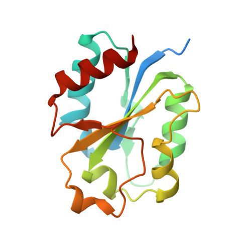
Molecular Mimicry in Innate Immunity: CRYSTAL STRUCTURE OF A BACTERIAL TIR DOMAIN.
Chan, S.L., Low, L.Y., Hsu, S., Li, S., Liu, T., Santelli, E., Le Negrate, G., Reed, J.C., Woods, V.L., Pascual, J.(2009) J Biol Chem 284: 21386-21392
- PubMed: 19535337
- DOI: https://doi.org/10.1074/jbc.C109.007591
- Primary Citation of Related Structures:
3H16 - PubMed Abstract:
Macrophages detect pathogen infection via the activation of their plasma membrane-bound Toll-like receptor proteins (TLRs). The heterotypic interaction between the Toll/interleukin-1 receptor (TIR) domains of TLRs and adaptor proteins, like Myeloid differentiation primary response gene 88 (MyD88), is the first intracellular step in the signaling pathway of the mammalian innate immune response. The hetero-oligomerization of the TIRs of the receptor and adaptor brings about the activation of the transcription factor NF-kappaB, which regulates the synthesis of pro-inflammatory cytokines. Here, we report the first crystal structure of a bacterial TIR domain solved at 2.5 A resolution. The three-dimensional fold of Paracoccus denitrificans TIR is identical to that observed for the TIR of human TLRs and MyD88 proteins. The structure shows a unique dimerization interface involving the DD-loop and EE-loop residues, whereas leaving the BB-loop highly exposed. Peptide amide hydrogen-deuterium exchange mass spectrometry also reveals that the same region is used for dimerization in solution and in the context of the full-length protein. These results, together with a functional interaction between P. denitrificans TIR and MyD88 visualized in a co-immunoprecipitation assay, further substantiate the model that bacterial TIR proteins adopt structural mimicry of the host active receptor TIR domains to interfere with the signaling of TLRs and their adaptors to decrease the inflammatory response.
Organizational Affiliation:
Inflammation and Infectious Diseases Center and Cancer Center, Burnham Institute for Medical Research, La Jolla, California 92037, USA.