Mechanisms of antagonism of the GluR2 AMPA receptor: structure and dynamics of the complex of two willardiine antagonists with the glutamate binding domain.
Ahmed, A.H., Thompson, M.D., Fenwick, M.K., Romero, B., Loh, A.P., Jane, D.E., Sondermann, H., Oswald, R.E.(2009) Biochemistry 48: 3894-3903
- PubMed: 19284741
- DOI: https://doi.org/10.1021/bi900107m
- Primary Citation of Related Structures:
3H03, 3H06 - PubMed Abstract:
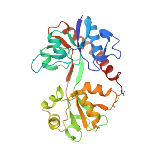
Ionotropic glutamate receptors mediate the majority of vertebrate excitatory synaptic transmission. The development of selective antagonists for glutamate receptor subtypes is of interest in the treatment of a variety of neurological disorders. This study presents the crystal structure of the binding domain of GluR2 bound to two antagonists (UBP277 and UBP282) that are derivatives of the natural product, willardiine. The antagonists bind to one lobe of the protein with interactions similar to agonists. Interaction with the second lobe differs between the two antagonists, resulting in a different position of the uracil ring and different orientations of the bilobed structure. UBP277 binding produces a stable lobe orientation that is similar to the apo state, but the binding of UBP282 produces the largest hyperextension of the lobes yet reported for an AMPA receptor. The carboxyethyl (UBP277) and carboxybenzyl (UBP282) substituents in the N(3) position keep the lobes separated by a "foot-in-the-door" mechanism and the internal dynamics are minimal compared to the CNQX-bound form of the protein (which makes minimal contacts with one of the two lobes). In contrast to the antagonists CNQX and DNQX, UBP277 and UBP282 produce complexes with higher thermal stability, but affinities that are more than 100-fold lower. These structures support the idea that antagonism is associated with the overall orientation of the lobes rather than with specific interactions, and antagonism can rise either from specific interactions with both lobes ("foot-in-the-door" mechanism) or from the lack of extensive interactions with one of the two lobes.
Organizational Affiliation:
Department of Molecular Medicine, Cornell University, Ithaca, New York 14853, USA.