3GJC

Crystal Structure of the E290S mutant of LeuT with bound OG


Experimental Data Snapshot

  • Method: X-RAY DIFFRACTION
  • Resolution: 2.80 Å
  • R-Value Free: 0.267 
  • R-Value Work: 0.221 
  • R-Value Observed: 0.223 

wwPDB Validation   3D Report Full Report


Ligand Structure Quality Assessment 


This is version 1.5 of the entry. See complete history


Literature

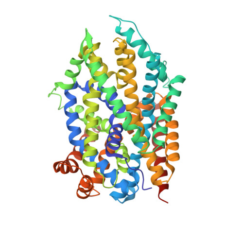
Binding of an octylglucoside detergent molecule in the second substrate (S2) site of LeuT establishes an inhibitor-bound conformation

Quick, M.Winther, A.M.L.Shi, L.Nissen, P.Weinstein, H.Javitch, J.A.

(2009) Proc Natl Acad Sci U S A 106: 5563-5568

  • DOI: https://doi.org/10.1073/pnas.0811322106
  • Primary Citation of Related Structures:  
    3GJC, 3GJD

  • PubMed Abstract: 

    The first crystal structure of the neurotransmitter/sodium symporter homolog LeuT revealed an occluded binding pocket containing leucine and 2 Na(+); later structures showed tricyclic antidepressants (TCAs) in an extracellular vestibule approximately 11 A above the bound leucine and 2 Na(+). We recently found this region to be a second binding (S2) site and that binding of substrate to this site triggers Na(+)-coupled substrate symport. Here, we show a profound inhibitory effect of n-octyl-beta-d-glucopyranoside (OG), the detergent used for LeuT crystallization, on substrate binding to the S2 site. In parallel, we determined at 2.8 A the structure of LeuT-E290S, a mutant that, like LeuT-WT, binds 2 substrate molecules. This structure was similar to that of WT and clearly revealed an OG molecule in the S2 site. We also observed electron density at the S2 site in LeuT-WT crystals, and this also was accounted for by an OG molecule in that site. Computational analyses, based on the available crystal structures of LeuT, indicated the nature of structural arrangements in the extracellular region of LeuT that differentiate the actions of substrates from inhibitors bound in the S2 site. We conclude that the current LeuT crystal structures, all of which have been solved in OG, represent functionally blocked forms of the transporter, whereas a substrate bound in the S2 site will promote a different state that is essential for Na(+)-coupled symport.


  • Organizational Affiliation

    Center for Molecular Recognition, Columbia University College of Physicians and Surgeons, 630 West 168th Street, New York, NY 10032, USA.


Macromolecules
Find similar proteins by:  (by identity cutoff)  |  3D Structure
Entity ID: 1
MoleculeChains Sequence LengthOrganismDetailsImage
Transporter
A, B
513Aquifex aeolicusMutation(s): 1 
Gene Names: aq_2077snf
Membrane Entity: Yes 
UniProt
Find proteins for O67854 (Aquifex aeolicus (strain VF5))
Explore O67854 
Go to UniProtKB:  O67854
Entity Groups  
Sequence Clusters30% Identity50% Identity70% Identity90% Identity95% Identity100% Identity
UniProt GroupO67854
Sequence Annotations
Expand
  • Reference Sequence
Small Molecules
Ligands 3 Unique
IDChains Name / Formula / InChI Key2D Diagram3D Interactions
BOG
Query on BOG

Download Ideal Coordinates CCD File 
E [auth A]
F [auth A]
G [auth A]
H [auth A]
I [auth B]
E [auth A],
F [auth A],
G [auth A],
H [auth A],
I [auth B],
L [auth B],
M [auth B],
N [auth B]
octyl beta-D-glucopyranoside
C14 H28 O6
HEGSGKPQLMEBJL-RKQHYHRCSA-N
LEU
Query on LEU

Download Ideal Coordinates CCD File 
D [auth A],
K [auth B]
LEUCINE
C6 H13 N O2
ROHFNLRQFUQHCH-YFKPBYRVSA-N
NA
Query on NA

Download Ideal Coordinates CCD File 
C [auth A],
J [auth B]
SODIUM ION
Na
FKNQFGJONOIPTF-UHFFFAOYSA-N
Experimental Data & Validation

Experimental Data

  • Method: X-RAY DIFFRACTION
  • Resolution: 2.80 Å
  • R-Value Free: 0.267 
  • R-Value Work: 0.221 
  • R-Value Observed: 0.223 
  • Space Group: P 1 21 1
Unit Cell:
Length ( Å )Angle ( ˚ )
a = 83.79α = 90
b = 93.51β = 94.22
c = 87.46γ = 90
Software Package:
Software NamePurpose
XSCALEdata scaling
PHASERphasing
PHENIXrefinement
PDB_EXTRACTdata extraction
MAR345data collection

Structure Validation

View Full Validation Report


Ligand Structure Quality Assessment 


Entry History 

Deposition Data

Revision History  (Full details and data files)

  • Version 1.0: 2009-04-28
    Type: Initial release
  • Version 1.1: 2011-07-13
    Changes: Version format compliance
  • Version 1.2: 2017-11-01
    Changes: Data collection, Refinement description
  • Version 1.3: 2020-07-29
    Type: Remediation
    Reason: Carbohydrate remediation
    Changes: Data collection, Derived calculations, Structure summary
  • Version 1.4: 2021-11-10
    Changes: Database references, Structure summary
  • Version 1.5: 2023-11-01
    Changes: Data collection, Refinement description