3GB4

Crystal Structure of Dicamba Monooxygenase with Non-heme Cobalt and Dicamba


Experimental Data Snapshot

  • Method: X-RAY DIFFRACTION
  • Resolution: 2.05 Å
  • R-Value Free: 0.262 
  • R-Value Work: 0.221 
  • R-Value Observed: 0.225 

wwPDB Validation   3D Report Full Report


Ligand Structure Quality Assessment 


This is version 1.5 of the entry. See complete history


Literature

Dicamba monooxygenase: structural insights into a dynamic Rieske oxygenase that catalyzes an exocyclic monooxygenation.

D'Ordine, R.L.Rydel, T.J.Storek, M.J.Sturman, E.J.Moshiri, F.Bartlett, R.K.Brown, G.R.Eilers, R.J.Dart, C.Qi, Y.Flasinski, S.Franklin, S.J.

(2009) J Mol Biol 392: 481-497

  • DOI: https://doi.org/10.1016/j.jmb.2009.07.022
  • Primary Citation of Related Structures:  
    3GB4, 3GOB, 3GTE, 3GTS, 6VSH

  • PubMed Abstract: 

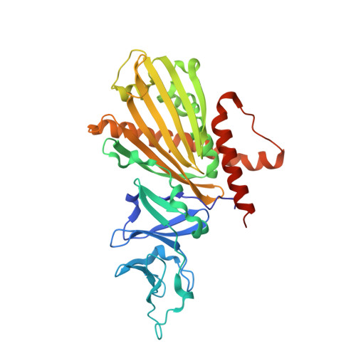
    Dicamba (2-methoxy-3,6-dichlorobenzoic acid) O-demethylase (DMO) is the terminal Rieske oxygenase of a three-component system that includes a ferredoxin and a reductase. It catalyzes the NADH-dependent oxidative demethylation of the broad leaf herbicide dicamba. DMO represents the first crystal structure of a Rieske non-heme iron oxygenase that performs an exocyclic monooxygenation, incorporating O(2) into a side-chain moiety and not a ring system. The structure reveals a 3-fold symmetric trimer (alpha(3)) in the crystallographic asymmetric unit with similar arrangement of neighboring inter-subunit Rieske domain and non-heme iron site enabling electron transport consistent with other structurally characterized Rieske oxygenases. While the Rieske domain is similar, differences are observed in the catalytic domain, which is smaller in sequence length than those described previously, yet possessing an active-site cavity of larger volume when compared to oxygenases with larger substrates. Consistent with the amphipathic substrate, the active site is designed to interact with both the carboxylate and aromatic ring with both key polar and hydrophobic interactions observed. DMO structures were solved with and without substrate (dicamba), product (3,6-dichlorosalicylic acid), and either cobalt or iron in the non-heme iron site. The substitution of cobalt for iron revealed an uncommon mode of non-heme iron binding trapped by the non-catalytic Co(2+), which, we postulate, may be transiently present in the native enzyme during the catalytic cycle. Thus, we present four DMO structures with resolutions ranging from 1.95 to 2.2 A, which, in sum, provide a snapshot of a dynamic enzyme where metal binding and substrate binding are coupled to observed structural changes in the non-heme iron and catalytic sites.


  • Organizational Affiliation

    Monsanto Company, Chesterfield MO 63017, USA. robert.l.d'ordine@monsanto.com


Macromolecules
Find similar proteins by:  (by identity cutoff)  |  3D Structure
Entity ID: 1
MoleculeChains Sequence LengthOrganismDetailsImage
DdmC
A, B, C
349Stenotrophomonas maltophiliaMutation(s): 1 
Gene Names: ddmC
UniProt
Find proteins for Q5S3I3 (Stenotrophomonas maltophilia)
Explore Q5S3I3 
Go to UniProtKB:  Q5S3I3
Entity Groups  
Sequence Clusters30% Identity50% Identity70% Identity90% Identity95% Identity100% Identity
UniProt GroupQ5S3I3
Sequence Annotations
Expand
  • Reference Sequence
Small Molecules
Ligands 3 Unique
IDChains Name / Formula / InChI Key2D Diagram3D Interactions
D3M
Query on D3M

Download Ideal Coordinates CCD File 
F [auth A],
J [auth B]
3,6-dichloro-2-methoxybenzoic acid
C8 H6 Cl2 O3
IWEDIXLBFLAXBO-UHFFFAOYSA-N
FES
Query on FES

Download Ideal Coordinates CCD File 
D [auth A],
H [auth B],
M [auth C]
FE2/S2 (INORGANIC) CLUSTER
Fe2 S2
NIXDOXVAJZFRNF-UHFFFAOYSA-N
CO
Query on CO

Download Ideal Coordinates CCD File 
E [auth A]
G [auth A]
I [auth B]
K [auth B]
L [auth B]
E [auth A],
G [auth A],
I [auth B],
K [auth B],
L [auth B],
N [auth C],
O [auth C]
COBALT (II) ION
Co
XLJKHNWPARRRJB-UHFFFAOYSA-N
Experimental Data & Validation

Experimental Data

  • Method: X-RAY DIFFRACTION
  • Resolution: 2.05 Å
  • R-Value Free: 0.262 
  • R-Value Work: 0.221 
  • R-Value Observed: 0.225 
  • Space Group: P 32
Unit Cell:
Length ( Å )Angle ( ˚ )
a = 81.55α = 90
b = 81.55β = 90
c = 161.29γ = 120
Software Package:
Software NamePurpose
SERGUIdata collection
PHASERphasing
CNXrefinement
HKL-2000data reduction
HKL-2000data scaling

Structure Validation

View Full Validation Report


Ligand Structure Quality Assessment 


Entry History 

Deposition Data

Revision History  (Full details and data files)

  • Version 1.0: 2009-07-28
    Type: Initial release
  • Version 1.1: 2011-07-13
    Changes: Version format compliance
  • Version 1.2: 2017-11-01
    Changes: Refinement description
  • Version 1.3: 2021-10-20
    Changes: Database references, Derived calculations
  • Version 1.4: 2024-02-21
    Changes: Data collection
  • Version 1.5: 2024-04-03
    Changes: Refinement description