3FYQ

Structure of Drosophila melanogaster talin IBS2 domain (residues 1981-2168)


Experimental Data Snapshot

  • Method: X-RAY DIFFRACTION
  • Resolution: 1.95 Å
  • R-Value Free: 0.255 
  • R-Value Work: 0.206 
  • R-Value Observed: 0.209 

wwPDB Validation   3D Report Full Report


This is version 1.2 of the entry. See complete history


Literature

Crystal structure of the talin integrin binding domain 2.

Cheung, T.Y.Fairchild, M.J.Zarivach, R.Tanentzapf, G.Van Petegem, F.

(2009) J Mol Biol 387: 787-793

  • DOI: https://doi.org/10.1016/j.jmb.2009.01.053
  • Primary Citation of Related Structures:  
    3FYQ

  • PubMed Abstract: 

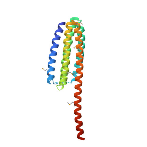
    Integrins are transmembrane receptors that mediate cell adhesion to the extracellular matrix and play essential roles in tissue development and maintenance. The cytoplasmic segment of integrin associates with talin, a large intracellular protein that links integrin to the actin cytoskeleton. Binding of talin via an integrin binding segment (IBS1) results in large conformational changes in the extracellular portion of integrin, which modulates the affinity of integrins for their extracellular matrix ligands. However, integrin binding also requires a second segment of talin (IBS2). Despite detailed descriptions of the integrin-IBS1 binding, the molecular determinants that drive the integrin-IBS2 association are poorly understood. Here, we describe the crystal structure of the talin IBS2 domain, which forms a five-helix bundle. The large structural homology with a vinculin binding domain hints at an ancient gene duplication and suggests that helix 4 may bind to vinculin if the bundle is unfolded. Mapping previous mutations on the surface highlights a likely binding interface for integrin.


  • Organizational Affiliation

    Department of Cellular and Physiological Sciences, University of British Columbia, Vancouver, Canada.


Macromolecules
Find similar proteins by:  (by identity cutoff)  |  3D Structure
Entity ID: 1
MoleculeChains Sequence LengthOrganismDetailsImage
CG6831-PA (Talin)199Drosophila melanogasterMutation(s): 0 
Gene Names: rheaCG6831Dmel_CG6831
UniProt
Find proteins for Q9VSL8 (Drosophila melanogaster)
Explore Q9VSL8 
Go to UniProtKB:  Q9VSL8
Entity Groups  
Sequence Clusters30% Identity50% Identity70% Identity90% Identity95% Identity100% Identity
UniProt GroupQ9VSL8
Sequence Annotations
Expand
  • Reference Sequence
Small Molecules
Modified Residues  1 Unique
IDChains TypeFormula2D DiagramParent
MSE
Query on MSE
A
L-PEPTIDE LINKINGC5 H11 N O2 SeMET
Experimental Data & Validation

Experimental Data

  • Method: X-RAY DIFFRACTION
  • Resolution: 1.95 Å
  • R-Value Free: 0.255 
  • R-Value Work: 0.206 
  • R-Value Observed: 0.209 
  • Space Group: C 1 2 1
Unit Cell:
Length ( Å )Angle ( ˚ )
a = 90.919α = 90
b = 40.079β = 112.87
c = 57.547γ = 90
Software Package:
Software NamePurpose
HKL-2000data collection
SHELXDphasing
REFMACrefinement
HKL-2000data reduction
HKL-2000data scaling

Structure Validation

View Full Validation Report


Entry History 

Deposition Data

Revision History  (Full details and data files)

  • Version 1.0: 2009-02-03
    Type: Initial release
  • Version 1.1: 2011-07-13
    Changes: Version format compliance
  • Version 1.2: 2018-01-24
    Changes: Structure summary