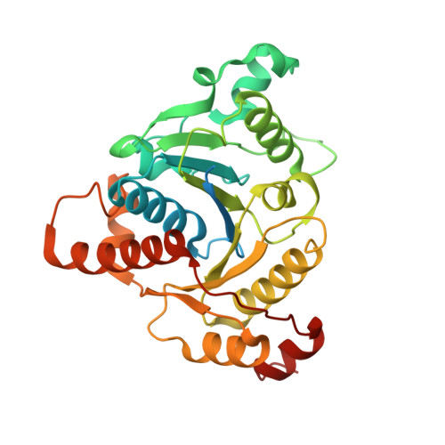
Crystal Structure of the L Protein of Rhodobacter sphaeroides Light-Independent Protochlorophyllide Reductase with MgADP Bound: A Homologue of the Nitrogenase Fe Protein.
Sarma, R., Barney, B.M., Hamilton, T.L., Jones, A., Seefeldt, L.C., Peters, J.W.(2008) Biochemistry 47: 13004-13015
- PubMed: 19006326
- DOI: https://doi.org/10.1021/bi801058r
- Primary Citation of Related Structures:
3FWY - PubMed Abstract:
The L protein (BchL) of the dark-operative protochlorophyllide reductase (DPOR) from Rhodobacter sphaeroides has been purified from an Azotobacter vinelandii expression system; its interaction with nucleotides has been examined, and the X-ray structure of the protein has been determined with bound MgADP to 1.6 A resolution. DPOR catalyzes the reduction of protochlorophyllide to chlorophyllide, a reaction critical to the biosynthesis of bacteriochlorophylls. The DPOR holoenzyme is comprised of two component proteins, the dimeric BchL protein and the heterotetrameric BchN/BchB protein. The DPOR component proteins share significant overall similarities with the nitrogenase Fe protein (NifH) and the MoFe (NifDK) protein, the enzyme system responsible for reduction of dinitrogen to ammonia. Here, BchL was expressed in A. vinelandii and purified to homogeneity using an engineered polyhistidine tag. The purified, recombinant BchL was found to contain 3.6 mol of Fe/mol of BchL homodimer, consistent with the presence of a [4Fe-4S] cluster and analogous to the [4Fe-4S] cluster present in the Fe protein. The MgATP- and MgADP-induced conformational changes in BchL were examined by an Fe chelation assay and found to be distinctly different from the nucleotide-stimulated Fe release observed for the Fe protein. The recombinant BchL was crystallized with bound MgADP, and the structure was determined to 1.6 A resolution. BchL is found to share overall structural similarity with the nitrogenase Fe protein, including the subunit bridging [4Fe-4S] cluster and nucleotide binding sites. Despite the high level of structural similarity, however, BchL is found to be incapable of substituting for the Fe protein in a nitrogenase substrate reduction assay. The newly determined structure of BchL and its comparison to its close homologue, the nitrogenase Fe protein, provide the basis for understanding how these highly related proteins can discriminate between their respective functions in microbial systems where each must function simultaneously.
Organizational Affiliation:
Department of Chemistry and Biochemistry and Astrobiology Biogeocatalysis Research Center, Montana State University, Bozeman, Montana 59717, USA.