3FHZ

Crystal structure of the arginine repressor from Mycobacterium tuberculosis bound with its DNA operator and co-repressor, L-arginine


Experimental Data Snapshot

  • Method: X-RAY DIFFRACTION
  • Resolution: 3.27 Å
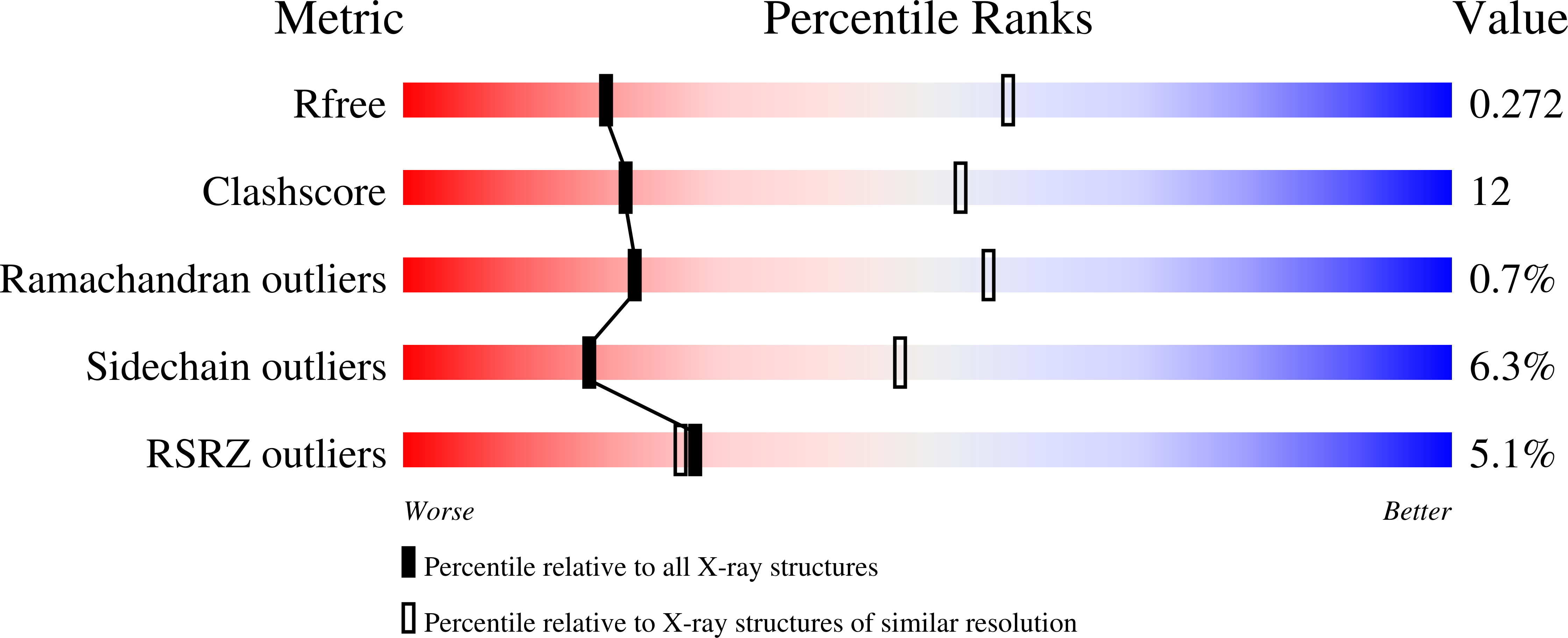
  • R-Value Free: 0.276 
  • R-Value Work: 0.231 
  • R-Value Observed: 0.233 

wwPDB Validation   3D Report Full Report


Ligand Structure Quality Assessment 


This is version 1.3 of the entry. See complete history


Literature

The structure of the arginine repressor from Mycobacterium tuberculosis bound with its DNA operator and Co-repressor, L-arginine.

Cherney, L.T.Cherney, M.M.Garen, C.R.James, M.N.

(2009) J Mol Biol 388: 85-97

  • DOI: https://doi.org/10.1016/j.jmb.2009.02.053
  • Primary Citation of Related Structures:  
    3FHZ

  • PubMed Abstract: 

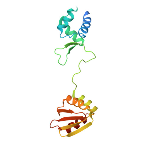
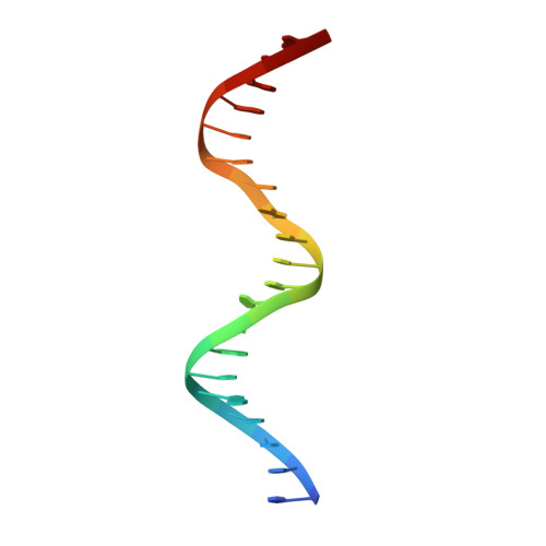
    The biosynthesis of arginine is an essential function for the metabolism of Mycobacterium tuberculosis (Mtb) and for the metabolism of many other microorganisms. The arginine repressor (ArgR) proteins control the transcription of genes encoding the arginine biosynthetic enzymes; they belong to repressors having one of the most intricate oligomerization patterns. Here, we present the crystal structure of the MtbArgR hexamer bound to three copies of the 20 base-pair DNA operator and to the co-repressor, L-arginine, determined to 3.3 A resolution. This is the first ternary structure of an intact hexameric ArgR in complex with its DNA operator. The structure reported here is very different from the suggested models of the ternary ArgR-DNA complexes; it has revealed the sophisticated symmetry of the complex and the presence of two remarkably different protomer conformations, folded and extended. Both features provide flexibility to DNA binding and are important for understanding the detailed function of ArgRs. Two of the 20 base-pair DNA operators align in a unified double-helical structure, suggesting the possible presence of a double ARG box in the promoter region of the Mtb arginine operon. Two pairs of protomers bind to the unified double ARG box so that the two folded protomers bind to the central half-sites of the double ARG box, whereas the two extended protomers bind to the remote half-sites. The protomers of the third pair bound to the single DNA operator also have a folded and an extended conformation. A probable mechanism for arginine repression is suggested on the basis of this structure.


  • Organizational Affiliation

    Group in Protein Structure and Function, Department of Biochemistry, University of Alberta, Edmonton, Alberta, Canada T6G 2H7.


Macromolecules

Find similar proteins by:  (by identity cutoff)  |  3D Structure
Entity ID: 1
MoleculeChains Sequence LengthOrganismDetailsImage
Arginine repressor
A, B, C, D, E
A, B, C, D, E, F
170Mycobacterium tuberculosis H37RvMutation(s): 0 
Gene Names: ahrCargRMT1695MTCY06H11.22Rv1657
UniProt
Find proteins for P9WPY9 (Mycobacterium tuberculosis (strain ATCC 25618 / H37Rv))
Explore P9WPY9 
Go to UniProtKB:  P9WPY9
Entity Groups  
Sequence Clusters30% Identity50% Identity70% Identity90% Identity95% Identity100% Identity
UniProt GroupP9WPY9
Sequence Annotations
Expand
  • Reference Sequence

Find similar nucleic acids by:  Sequence   |   3D Structure  

Entity ID: 2
MoleculeChains LengthOrganismImage
5'-D(*TP*GP*TP*TP*GP*CP*AP*TP*AP*AP*CP*GP*AP*TP*GP*CP*AP*AP*AP*A)-3'
G, I, K
20N/A
Sequence Annotations
Expand
  • Reference Sequence

Find similar nucleic acids by:  Sequence   |   3D Structure  

Entity ID: 3
MoleculeChains LengthOrganismImage
5'-D(*TP*TP*TP*TP*GP*CP*AP*TP*CP*GP*TP*TP*AP*TP*GP*CP*AP*AP*CP*A)-3'
H, J, L
20N/A
Sequence Annotations
Expand
  • Reference Sequence
Small Molecules
Ligands 3 Unique
IDChains Name / Formula / InChI Key2D Diagram3D Interactions
ARG
Query on ARG

Download Ideal Coordinates CCD File 
M [auth A]
N [auth A]
P [auth B]
Q [auth B]
S [auth C]
M [auth A],
N [auth A],
P [auth B],
Q [auth B],
S [auth C],
T [auth C],
U [auth D],
W [auth E],
X [auth F]
ARGININE
C6 H15 N4 O2
ODKSFYDXXFIFQN-BYPYZUCNSA-O
GOL
Query on GOL

Download Ideal Coordinates CCD File 
V [auth D]GLYCEROL
C3 H8 O3
PEDCQBHIVMGVHV-UHFFFAOYSA-N
ACT
Query on ACT

Download Ideal Coordinates CCD File 
O [auth A],
R [auth B]
ACETATE ION
C2 H3 O2
QTBSBXVTEAMEQO-UHFFFAOYSA-M
Experimental Data & Validation

Experimental Data

  • Method: X-RAY DIFFRACTION
  • Resolution: 3.27 Å
  • R-Value Free: 0.276 
  • R-Value Work: 0.231 
  • R-Value Observed: 0.233 
  • Space Group: P 21 21 21
Unit Cell:
Length ( Å )Angle ( ˚ )
a = 63.286α = 90
b = 152.649β = 90
c = 163.423γ = 90
Software Package:
Software NamePurpose
MAR345data collection
MOLREPphasing
PHENIXrefinement
HKL-2000data reduction
HKL-2000data scaling

Structure Validation

View Full Validation Report


Ligand Structure Quality Assessment 


Entry History 

Deposition Data

Revision History  (Full details and data files)

  • Version 1.0: 2009-03-24
    Type: Initial release
  • Version 1.1: 2011-07-13
    Changes: Non-polymer description, Version format compliance
  • Version 1.2: 2017-11-01
    Changes: Refinement description
  • Version 1.3: 2023-09-06
    Changes: Data collection, Database references, Derived calculations, Refinement description