3FGC

Crystal Structure of the Bacterial Luciferase:Flavin Complex Reveals the Basis of Intersubunit Communication


Experimental Data Snapshot

  • Method: X-RAY DIFFRACTION
  • Resolution: 2.30 Å
  • R-Value Free: 0.241 
  • R-Value Work: 0.184 
  • R-Value Observed: 0.187 

wwPDB Validation   3D Report Full Report


Ligand Structure Quality Assessment 


This is version 1.3 of the entry. See complete history


Literature

Crystal structure of the bacterial luciferase/flavin complex provides insight into the function of the beta subunit.

Campbell, Z.T.Weichsel, A.Montfort, W.R.Baldwin, T.O.

(2009) Biochemistry 48: 6085-6094

  • DOI: https://doi.org/10.1021/bi900003t
  • Primary Citation of Related Structures:  
    3FGC

  • PubMed Abstract: 

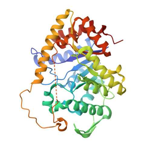
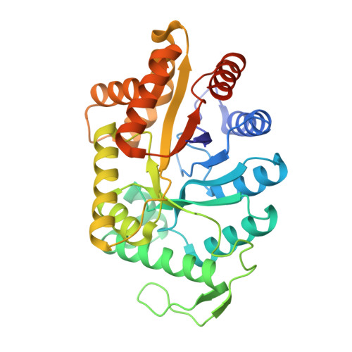
    Bacterial luciferase from Vibrio harveyi is a heterodimer composed of a catalytic alpha subunit and a homologous but noncatalytic beta subunit. Despite decades of enzymological investigation, structural evidence defining the active center has been elusive. We report here the crystal structure of V. harveyi luciferase bound to flavin mononucleotide (FMN) at 2.3 A. The isoalloxazine ring is coordinated by an unusual cis-Ala-Ala peptide bond. The reactive sulfhydryl group of Cys106 projects toward position C-4a, the site of flavin oxygenation. This structure also provides the first data specifying the conformations of a mobile loop that is crystallographically disordered in both prior crystal structures [(1995) Biochemistry 34, 6581-6586; (1996) J. Biol. Chem. 271, 21956 21968]. This loop appears to be a boundary between solvent and the active center. Within this portion of the protein, a single contact was observed between Phe272 of the alpha subunit, not seen in the previous structures, and Tyr151 of the beta subunit. Substitutions at position 151 on the beta subunit caused reductions in activity and total quantum yield. Several of these mutants were found to have decreased affinity for reduced flavin mononucleotide (FMNH(2)). These findings partially address the long-standing question of how the beta subunit stabilizes the active conformation of the alpha subunit, thereby participating in the catalytic mechanism.


  • Organizational Affiliation

    Department of Biochemistry and Molecular Biophysics, University of Arizona, 1041 East Lowell Street, Biological Sciences West, Tucson, Arizona 85721-0088, USA.


Macromolecules
Find similar proteins by:  (by identity cutoff)  |  3D Structure
Entity ID: 1
MoleculeChains Sequence LengthOrganismDetailsImage
Alkanal monooxygenase alpha chain
A, C
355Vibrio harveyiMutation(s): 0 
Gene Names: luxA
EC: 1.14.14.3
UniProt
Find proteins for P07740 (Vibrio harveyi)
Explore P07740 
Go to UniProtKB:  P07740
Entity Groups  
Sequence Clusters30% Identity50% Identity70% Identity90% Identity95% Identity100% Identity
UniProt GroupP07740
Sequence Annotations
Expand
  • Reference Sequence
Find similar proteins by:  (by identity cutoff)  |  3D Structure
Entity ID: 2
MoleculeChains Sequence LengthOrganismDetailsImage
Alkanal monooxygenase beta chain
B, D
332Vibrio harveyiMutation(s): 0 
Gene Names: luxB
EC: 1.14.14.3
UniProt
Find proteins for P07739 (Vibrio harveyi)
Explore P07739 
Go to UniProtKB:  P07739
Entity Groups  
Sequence Clusters30% Identity50% Identity70% Identity90% Identity95% Identity100% Identity
UniProt GroupP07739
Sequence Annotations
Expand
  • Reference Sequence
Small Molecules
Ligands 3 Unique
IDChains Name / Formula / InChI Key2D Diagram3D Interactions
FMN
Query on FMN

Download Ideal Coordinates CCD File 
E [auth A]FLAVIN MONONUCLEOTIDE
C17 H21 N4 O9 P
FVTCRASFADXXNN-SCRDCRAPSA-N
SO4
Query on SO4

Download Ideal Coordinates CCD File 
G [auth A]
H [auth A]
I [auth A]
J [auth A]
K [auth A]
G [auth A],
H [auth A],
I [auth A],
J [auth A],
K [auth A],
L [auth B],
M [auth B],
N [auth B],
O [auth C],
P [auth C],
Q [auth C],
S [auth D],
T [auth D],
U [auth D],
V [auth D],
W [auth D]
SULFATE ION
O4 S
QAOWNCQODCNURD-UHFFFAOYSA-L
PO4
Query on PO4

Download Ideal Coordinates CCD File 
F [auth A],
R [auth C]
PHOSPHATE ION
O4 P
NBIIXXVUZAFLBC-UHFFFAOYSA-K
Binding Affinity Annotations 
IDSourceBinding Affinity
FMN PDBBind:  3FGC Kd: 1.20e+5 (nM) from 1 assay(s)
Experimental Data & Validation

Experimental Data

  • Method: X-RAY DIFFRACTION
  • Resolution: 2.30 Å
  • R-Value Free: 0.241 
  • R-Value Work: 0.184 
  • R-Value Observed: 0.187 
  • Space Group: P 21 21 21
Unit Cell:
Length ( Å )Angle ( ˚ )
a = 58.886α = 90
b = 109.301β = 90
c = 301.419γ = 90
Software Package:
Software NamePurpose
HKL-2000data collection
PHASERphasing
REFMACrefinement
DENZOdata reduction
SCALEPACKdata scaling

Structure Validation

View Full Validation Report


Ligand Structure Quality Assessment 


Entry History 

Deposition Data

Revision History  (Full details and data files)

  • Version 1.0: 2009-05-26
    Type: Initial release
  • Version 1.1: 2011-07-13
    Changes: Version format compliance
  • Version 1.2: 2017-11-01
    Changes: Refinement description
  • Version 1.3: 2023-09-06
    Changes: Data collection, Database references, Derived calculations, Refinement description