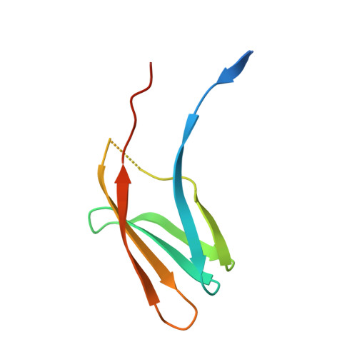
Caf1A usher possesses a Caf1 subunit-like domain that is crucial for Caf1 fibre secretion
Yu, X., Visweswaran, G.R., Duck, Z., Marupakula, S., MacIntyre, S., Knight, S.D., Zavialov, A.V.(2009) Biochem J 418: 541-551
- PubMed: 19032149
- DOI: https://doi.org/10.1042/BJ20080992
- Primary Citation of Related Structures:
3FCG - PubMed Abstract:
The chaperone/usher pathway controls assembly of fibres of adhesive organelles of Gram-negative bacteria. The final steps of fibre assembly and fibre translocation to the cell surface are co-ordinated by the outer membrane proteins, ushers. Ushers consist of several soluble periplasmic domains and a single transmembrane beta-barrel. Here we report isolation and structural/functional characterization of a novel middle domain of the Caf1A usher from Yersinia pestis. The isolated UMD (usher middle domain) is a highly soluble monomeric protein capable of autonomous folding. A 2.8 A (1 A=0.1 nm) resolution crystal structure of UMD revealed that this domain has an immunoglobulin-like fold similar to that of donor-strand-complemented Caf1 fibre subunit. Moreover, these proteins displayed significant structural similarity. Although UMD is in the middle of the predicted amphipathic beta-barrel of Caf1A, the usher still assembled in the membrane in the absence of this domain. UMD did not bind Caf1M-Caf1 complexes, but its presence was shown to be essential for Caf1 fibre secretion. The study suggests that UMD may play the role of a subunit-substituting protein (dummy subunit), plugging or priming secretion through the channel in the Caf1A usher. Comparison of isolated UMD with the recent structure of the corresponding domain of PapC usher revealed high similarity of the core structures, suggesting a universal structural adaptation of FGL (F(1)G(1) long) and FGS (F(1)G(1) short) chaperone/usher pathways for the secretion of different types of fibres. The functional role of two topologically different states of this plug domain suggested by structural and biochemical results is discussed.
Organizational Affiliation:
Department of Molecular Biology, Uppsala Biomedical Centre, Swedish University of Agricultural Sciences, Uppsala, Sweden.