3F8D

Structure of Sulfolobus solfataricus Thioredoxin reductase Mutant C147A


Experimental Data Snapshot

  • Method: X-RAY DIFFRACTION
  • Resolution: 1.40 Å
  • R-Value Free: 0.226 
  • R-Value Work: 0.206 
  • R-Value Observed: 0.209 

wwPDB Validation   3D Report Full Report


Ligand Structure Quality Assessment 


This is version 1.3 of the entry. See complete history


Literature

Structure and stability of a thioredoxin reductase from Sulfolobus solfataricus: a thermostable protein with two functions

Ruggiero, A.Masullo, M.Ruocco, M.R.Grimaldi, P.Lanzotti, M.A.Arcari, P.Zagari, A.Vitagliano, L.

(2009) Biochim Biophys Acta 1794: 554-562

  • DOI: https://doi.org/10.1016/j.bbapap.2008.11.011
  • Primary Citation of Related Structures:  
    3F8D, 3F8P, 3F8R

  • PubMed Abstract: 

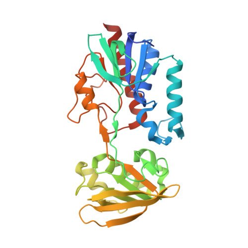
    Recent investigations have demonstrated that disulfide bridges may play a crucial role in the stabilization of proteins in hyperthermophilic organisms. A major role in the process of disulfide formation is played by ubiquitous proteins belonging to the thioredoxin superfamily, which includes thioredoxins (Trx), thioredoxin reductases (TrxR), and disulfide oxidases/isomerases (PDO/PDI). Here we report a characterization of the structure and stability of the TrxR (SsTrxRB3) isolated from the archaeon Sulfolobus solfataricus. This protein is particularly interesting since it is able to process different substrates (Trxs and PDO) and it is endowed with an additional NADH oxidase activity. The crystal structure of the wild-type enzyme, of its complex with NADP and of the C147A mutant provides interesting clues on the enzyme function. In contrast to what is observed for class II TrxRs, in the structure of the oxidized enzyme, the FAD binding site is occupied by a partially disordered NAD molecule. In the active site of the C147A mutant, which exhibits a marginal NADH oxidase activity, the FAD is canonically bound to the enzyme. Molecular modeling indicates that a FAD molecule can be accommodated in the site of the reduced SsTrxRB3. Depending on the oxidation state, SsTrxRB3 can bind a different cofactor in its active site. This peculiar feature has been related to its dual activity. Denaturation experiments followed by circular dichroism indicate that electrostatic interactions play an important role in the stabilization of this thermostable protein. The analysis of the enzyme 3D-structure has also provided insights into the bases of SsTrxRB3 stability.


  • Organizational Affiliation

    Istituto di Biostrutture e Bioimmagini, CNR, Via Mezzocannone 16, I-80134 Napoli, Italy.


Macromolecules
Find similar proteins by:  (by identity cutoff)  |  3D Structure
Entity ID: 1
MoleculeChains Sequence LengthOrganismDetailsImage
Thioredoxin reductase (TrxB-3)
A, B, C, D
323Saccharolobus solfataricusMutation(s): 1 
EC: 1.6.4.5
UniProt
Find proteins for Q8X236 (Saccharolobus solfataricus)
Explore Q8X236 
Go to UniProtKB:  Q8X236
Entity Groups  
Sequence Clusters30% Identity50% Identity70% Identity90% Identity95% Identity100% Identity
UniProt GroupQ8X236
Sequence Annotations
Expand
  • Reference Sequence
Small Molecules
Ligands 3 Unique
IDChains Name / Formula / InChI Key2D Diagram3D Interactions
FAD
Query on FAD

Download Ideal Coordinates CCD File 
E [auth A],
I [auth B],
L [auth C],
N [auth D]
FLAVIN-ADENINE DINUCLEOTIDE
C27 H33 N9 O15 P2
VWWQXMAJTJZDQX-UYBVJOGSSA-N
SO4
Query on SO4

Download Ideal Coordinates CCD File 
F [auth A]
G [auth A]
J [auth B]
K [auth B]
M [auth C]
F [auth A],
G [auth A],
J [auth B],
K [auth B],
M [auth C],
O [auth D]
SULFATE ION
O4 S
QAOWNCQODCNURD-UHFFFAOYSA-L
ACY
Query on ACY

Download Ideal Coordinates CCD File 
H [auth B]ACETIC ACID
C2 H4 O2
QTBSBXVTEAMEQO-UHFFFAOYSA-N
Experimental Data & Validation

Experimental Data

  • Method: X-RAY DIFFRACTION
  • Resolution: 1.40 Å
  • R-Value Free: 0.226 
  • R-Value Work: 0.206 
  • R-Value Observed: 0.209 
  • Space Group: P 21 21 21
Unit Cell:
Length ( Å )Angle ( ˚ )
a = 76.581α = 90
b = 121.874β = 90
c = 126.843γ = 90
Software Package:
Software NamePurpose
ADSCdata collection
AMoREphasing
CNSrefinement
DENZOdata reduction
SCALEPACKdata scaling

Structure Validation

View Full Validation Report


Ligand Structure Quality Assessment 


Entry History 

Deposition Data

Revision History  (Full details and data files)

  • Version 1.0: 2009-01-13
    Type: Initial release
  • Version 1.1: 2011-07-13
    Changes: Version format compliance
  • Version 1.2: 2021-11-10
    Changes: Database references
  • Version 1.3: 2023-11-01
    Changes: Data collection, Refinement description