3EMV

Crystal structure of Plasmodium vivax PNP with sulphate


Experimental Data Snapshot

  • Method: X-RAY DIFFRACTION
  • Resolution: 1.85 Å
  • R-Value Free: 0.233 
  • R-Value Work: 0.193 
  • R-Value Observed: 0.195 

wwPDB Validation   3D Report Full Report


This is version 1.2 of the entry. See complete history


Literature

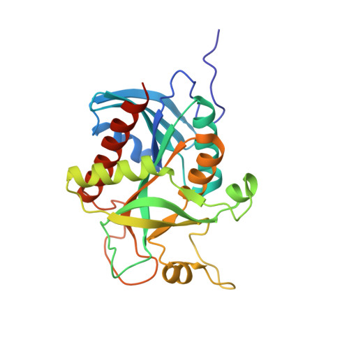
Conservation of structure and activity in Plasmodium purine nucleoside phosphorylases

Chaikuad, A.Brady, R.L.

(2009) BMC Struct Biol 9: 42-42

  • DOI: https://doi.org/10.1186/1472-6807-9-42
  • Primary Citation of Related Structures:  
    3EMV, 3ENZ

  • PubMed Abstract: 

    Purine nucleoside phosphorylase (PNP) is central to purine salvage mechanisms in Plasmodium parasites, the causative agents of malaria. Most human malaria results from infection either by Plasmodium falciparum (Pf), the deadliest form of the parasite, or by the widespread Plasmodium vivax (Pv). Whereas the PNP enzyme from Pf has previously been studied in detail, despite the prevalence of Pv little is known about many of the key metabolic enzymes from this parasite, including PvPNP.


  • Organizational Affiliation

    Department of Biochemistry, University of Bristol, Bristol, BS8 1TD, UK. Apirat.Chaikuad@sgc.ox.ac.uk


Macromolecules
Find similar proteins by:  (by identity cutoff)  |  3D Structure
Entity ID: 1
MoleculeChains Sequence LengthOrganismDetailsImage
Uridine phosphorylase, putative253Plasmodium vivaxMutation(s): 0 
Gene Names: PVX_080575
EC: 2.4.2.1
UniProt
Find proteins for A5K9M4 (Plasmodium vivax (strain Salvador I))
Explore A5K9M4 
Go to UniProtKB:  A5K9M4
Entity Groups  
Sequence Clusters30% Identity50% Identity70% Identity90% Identity95% Identity100% Identity
UniProt GroupA5K9M4
Sequence Annotations
Expand
  • Reference Sequence
Small Molecules
Ligands 1 Unique
IDChains Name / Formula / InChI Key2D Diagram3D Interactions
SO4
Query on SO4

Download Ideal Coordinates CCD File 
B [auth A]SULFATE ION
O4 S
QAOWNCQODCNURD-UHFFFAOYSA-L
Experimental Data & Validation

Experimental Data

  • Method: X-RAY DIFFRACTION
  • Resolution: 1.85 Å
  • R-Value Free: 0.233 
  • R-Value Work: 0.193 
  • R-Value Observed: 0.195 
  • Space Group: H 3 2
Unit Cell:
Length ( Å )Angle ( ˚ )
a = 94.749α = 90
b = 94.749β = 90
c = 121.254γ = 120
Software Package:
Software NamePurpose
PHASERphasing
REFMACrefinement
HKL-2000data reduction
HKL-2000data scaling

Structure Validation

View Full Validation Report


Entry History 

Deposition Data

Revision History  (Full details and data files)

  • Version 1.0: 2009-08-04
    Type: Initial release
  • Version 1.1: 2011-07-13
    Changes: Version format compliance
  • Version 1.2: 2023-11-01
    Changes: Data collection, Database references, Derived calculations, Refinement description